Anti-Mouse CD172a - Purified in vivo GOLD™ Functional Grade
Antibody Clonality:
Monoclonal
Applications:- Blocking
- Flow Cytometry
- Immunohistochemistry- Frozen Section (IHC-F)
- Immunoprecipitation (IP)
- In Vivo Assay
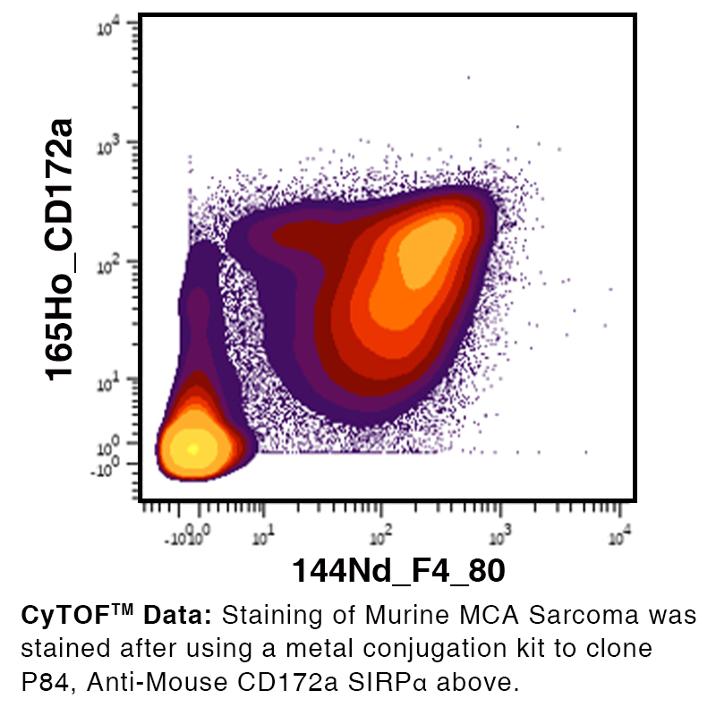
- Mass Cytometry (CyTOF)
Storage:
Functional grade preclinical antibodies may be stored sterile as received at 2-8°C for up to one month. For longer term storage aseptically aliquot in working volumes without diluting and store at -80°C. Avoid Repeated Freeze Thaw Cycles.
No additional charges, what you see is what you pay! *
Stay in control of your spending. These prices have no additional charges, not even shipping!
* Rare exceptions are clearly labelled (only 0.14% of items!).
Multibuy discounts available!
Contact us to find what you can save.
This product comes from:
US.
Typical lead time:
14-21 working days.
Contact us for more accurate information.
- Further Information
- References
- Related Products
- Show All
Further Information
CD172a is expressed on monocytes, macrophages, dendritic cells, and neuronal cells.
? 5.0 mg/ml
in vivo GOLD™, Purified in vivo Functional Grade
This monoclonal antibody is aseptically packaged and formulated in 0.01 M phosphate buffered saline (150 mM NaCl) PBS pH 7.2 - 7.4 with no carrier protein, potassium, calcium or preservatives added. Due to inherent biochemical properties of antibodies, certain products may be prone to precipitation over time. Precipitation may be removed by aseptic centrifugation and/or filtration.
This monoclonal antibody is aseptically packaged and formulated in 0.01 M phosphate buffered saline (150 mM NaCl) PBS pH 7.2 - 7.4 with no carrier protein, potassium, calcium or preservatives added. Due to inherent biochemical properties of antibodies, certain products may be prone to precipitation over time. Precipitation may be removed by aseptic centrifugation and/or filtration.
Mouse brain membrane protein
CD172a antibody, clone P84, recognizes CD172a, also known as single regulatory protein α (SIRPα) (signal regulatory protein alpha) or Src homology 2 domain-containing phosphatase substrate-1 (SHP-1), a type I transmembrane glycoprotein with three Ig-like extracellular domains and two cytoplasmic immunoreceptor tyrosine-based inhibition motifs (ITIMs)1. SIRPα is expressed predominantly in myeloid cells2 - including monocytes, macrophages, and dendritic cells (DCs) - and neuronal cells3. The extracellular ligand for SIRPα, CD47 (or integrin-associated protein [IAP])4, is expressed in most cell types5. In macrophages, ligation of SIRPα by CD47 inhibits macrophage phagocytosis of self cells6,7. SIRPα also negatively regulates DC-mediated T cell activation and DC maturation8-10. CD47 is also upregulated on tumor cells, inhibiting the phagocytosis of tumor cells by macrophages11. Therapeutics targeting the CD47-SIRPα interaction, including antibodies and fusion proteins, are currently under preclinical and clinical study for various malignancies as a monotherapy or in combination with other therapeutics12.
19261
?95% monomer by analytical SEC, >95% by SDS Page
CD172a