
Mitophagy is the selective degradation of old or depolarised mitochondria by autophagy and contributes to maintaining a healthy population of mitochondria. Since damaged mitochondria lead to collapse cell homoeostasis, mitophagy is believed to be protective against diseases related to mitochondrial dysfunction such as in neurodegenerative disorders.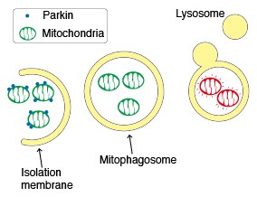
Parkin, an ubiquitin ligase known as the gene responsible for Parkinson’s disease, plays an important role in the autophagic elimination of mitophagy. When mitochondria are depolarised and dysfunctional, PTEN-induced putative kinase protein 1 (PINK1) accumulates on the outer membrane and recruits Parkin on the damaged mitochondria. The outer membrane on the mitochondria is then ubiquitinated through the ubiquitin ligase activity of Parkin. Finally, the poly-ubiquitinated mitochondria are selectively recognised and executed by the autophagic process.
Emission wavelength character of Keima-Red under various pH condition
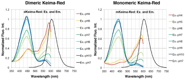
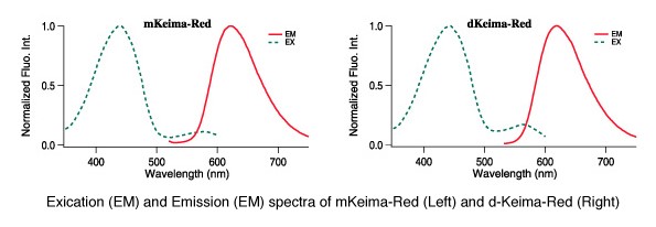
The fluorescent protein Keima has an excitation spectrum that changes according to pH. A short wavelength (440 nm) is predominant for excitation in a neutral environment, whereas a long wavelength (586 nm) is predominant in an acidic environment. The ratio of fluorescent intensity in each excitation condition is an indicator of mitophagy in living cells. CoralHue® dimeric Keima-Red (dKeima-Red) and CoralHue® monomeric Keima-Red (mKeima-Red) are red fluorescent proteins with extremely large Stokes shift. They absorb light maximally at 440 nm and emit red light at 616 nm and 620 nm, respectively. There are no other fluorescent proteins with this unique fluorescence. Due of this characteristic, they are excited by a very short wavelength but emit a long wavelength. The large Stokes shift property of Keima-Red allows effective applications such as for single wavelength excitation simultaneous multi-color imaging and single laser line FCCS.
Mitochondrial targeting-mKeima-Red for mitophagy detection
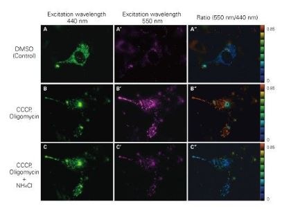 When MT-mKeima-Red, which is the Keima tagged with a mitochondrial-localised signal peptide sequence, and Parkin is expressed in target cells, mitophagy is detected and is visualised through the difference in the fluorescent wavelengths observed before and after drug treatment. Mitophagy is induced by the administration of CCCP and oligomycin, drugs that affect mitochondrial membrane potential and is observed with 550nm/440nm ratio images (A”, B”, C”).
Through mitophagy induction, the mitochondria-localised MT-mKeima-Red is displayed in red in ratio images showing its localisation in an acidic environment (B-B”). The neutraliser NH4Cl is then administered which forces the entire cell into a neutral environment and the image turns to blue (C-C”). The result matched findings in which the progression of mitophagy causes mitochondria to be engulfed in lysosomes when in an acidic environment.
Products
| Product Code | Description | Target | Size | Product Type |
|---|---|---|---|---|
| M126-3B | Anti-Keima-Red Trial Size | Keima-Red | 10 μg | Antibody |
| M127-3B | Anti-Keima-Red Trial Size | Keima-Red | 10 μg | Antibody |
| M127-3M | Anti-Keima-Red mAb | Keima-Red | 100 μg | Antibody |
| M126-3M | Anti-monomeric Keima-Red mAb | Keima-Red | 100 µL | Antibody |
| M182-3M | Anti-Keima-Red mAb | Keima | 100 μl | Antibody |
| AM-V0251 | CoralHue® Mitochondria-targeted mKeima-Red Expression Plasmid (pMT-mKeima-Red) | Mitochondria-targeted mKeima-Red | 20 µg | Vector |
| M182-3B | Anti-Keima Monoclonal Antibody Trial Size | Keima | 10 μg | Antibody |
| AM-V0091 | CoralHue® Monomeric Keima-Red (pmKeima-Red-S1) | Keima Red-pm Keima-Red-S1 | 20 µg | Vector |
| AM-V0101 | CoralHue® Dimeric Keima-Red (pdKeima-Red-S1) | Keima Red-pdKeima-Red-S1 | 20 µg | Vector |
| AM-V0104 | CoralHue® Humanized-codon dimeric Keima-Red (phdKeima-Red-S1) | Keima-Red (phdKeima-Red-S1) | 20 µg | Vector |
| AM-V0094 | CoralHue® Humanized-codon monomeric Keima-Red (phmKeima-Red-S1) | Keima-Red (phmKeima-Red-S1) | 20 µg | Vector |
| AM-V0093 | CoralHue® Monomeric Keima-Red (pmKeima-Red-MN1) | Keima Redpm Keima-Red-MN1 | 20 µg | Vector |
| AM-VS0304 | CoralHue® Monomer/Wild-type Comparison Set-Keima-Red (pmKeima-Red-S+pdKeima-Red-S1) | Monomer/Wild-type Comparison Set-Keima-Red (pmKeima-Red-S+pdKeima-Red-S1) | 2 x 20 μg | Vector |
| AM-V0090 | CoralHue® Humanized-codon monomeric Keima-Red (phmKeima-Red-MNLinker) | Keima-Red (phmKeima-Red-MNLinker) | 20 µg | Vector |
| AM-V0109 | CoralHue® Humanized-codon dimeric Keima-Red (phdKeima-Red-MCLinker) | Keima-Red (phdKeima-Red-MCLinker) | 20 µg | Vector |
| AM-V0099 | CoralHue® Humanized-codon monomeric Keima-Red (phmKeima-Red-MCLinker) | Keima-Red (phmKeima-Red-MCLinker) | 20 µg | Vector |
| AM-V0100 | CoralHue® Humanized-codon dimeric Keima-Red (phdKeima-Red-MNLinker) | Keima-Red (phdKeima-Red-MNLinker) | 20 µg | Vector |
| AM-V0274 | CoralHue® Nucleoplasm-targeted Humanized-codon dimeric Keima-Red Expression Plasmid (pNP-hdKeima-Red) | pNP-hdKeima-Red | 20 µg | Vector |
| AM-V0253 | CoralHue® Plasma Membrane-targeted mKeima-Red Expression Plasmid (pPM-mKeima-Red) | Plasma Membrane-targeted mKeima-Red | 20 µg | Vector |
| AM-V0124 | CoralHue® Humanized-codon dimeric Keima570 (phdKeima570-S1) | Keima 570(phdKeima570-S1) | 20 µg | Vector |
| AM-V0324 | CoralHue® Nucleoplasm-targeted Humanized-codon dimeric Keima570 Expression Plasmid (pNP-hdKeima570) | Nucleoplasm-targeted humanized-codon dimeric Keima570 (pNP-hdKeima570) | 20 µg | Vector |
For more information about this product range, get in touch.