LAG3 Antibody [5F11]
| Code | Size | Price |
|---|
| PSI-RF16088-0.02mg | 0.02mg | £150.00 |
Quantity:
| PSI-RF16088-0.1mg | 0.1mg | £515.00 |
Quantity:
Prices exclude any Taxes / VAT
Overview
Host Type: Mouse
Antibody Isotype: IgG1,k
Antibody Clonality: Monoclonal
Antibody Clone: 5F11
Regulatory Status: RUO
Target Species: Human
Applications:
- Immunocytochemistry (ICC)
- Immunofluorescence (IF)
- Immunohistochemistry- Paraffin Embedded (IHC-P)
- Western Blot (WB)
Shipping:
blue ice
Storage:
LAG-3 antibody can be stored at 4˚C for three months and -20˚C, stable for up to one year. As with all antibodies care should be taken to avoid repeated freeze thaw cycles. Antibodies should not be exposed to prolonged high temperatures.
Images
Documents
Further Information
Additional Names:
LAG3 Antibody: lymphocyte activating 3, LAG-3, CD223
Application Note:
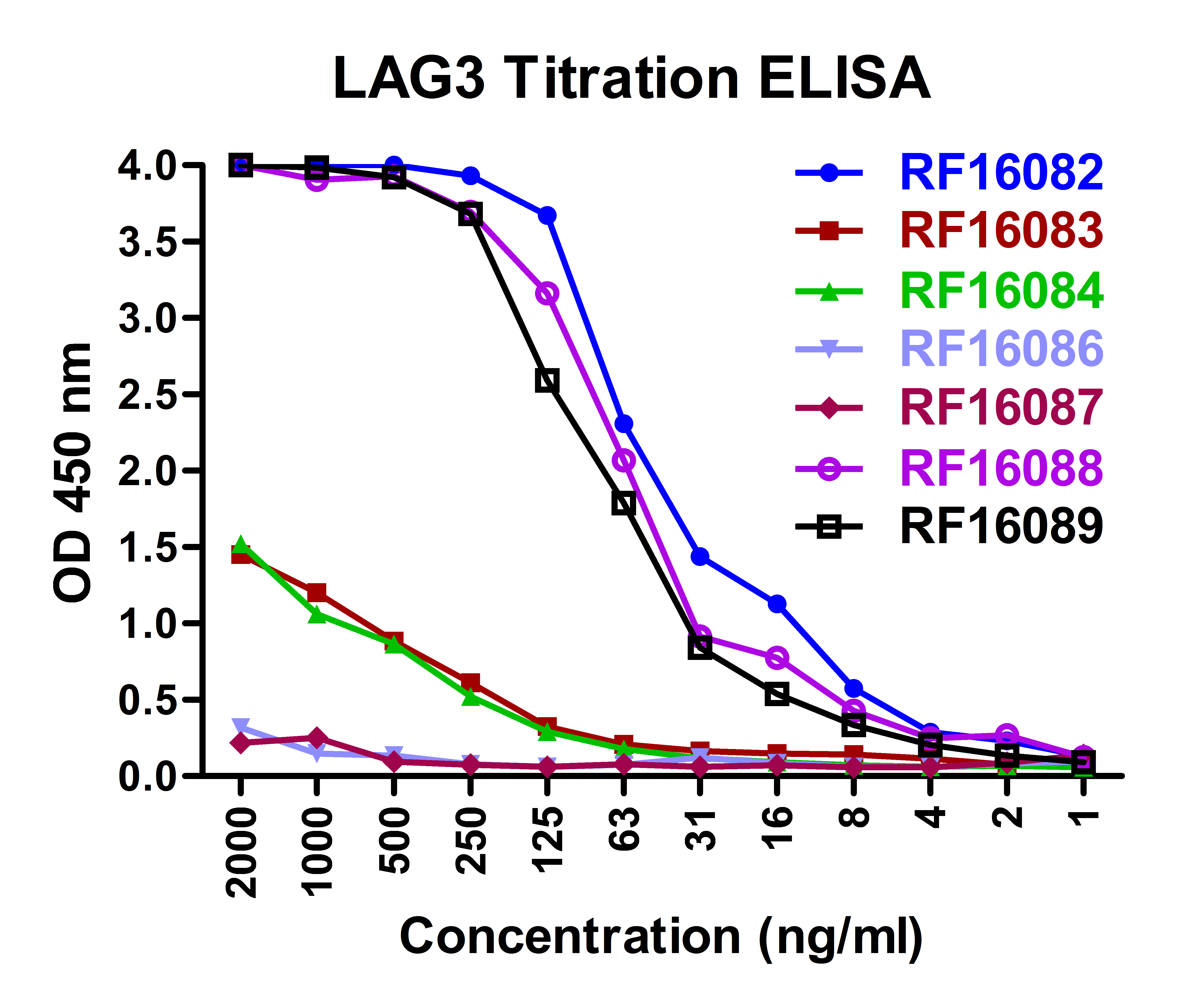
LAG-3 antibody can be used for ELISA starting at 0.25 μg/mL. For Western blot start at 0.25 μg/mL. For Immunocytochemistry start at 1 μg/mL. For Immunofluorescence start at 10 μg/mL. For Immunohistochemistry start at 5 μg/mL. For Flow Cytometry start at 1 μg/ml.
Antibody validated: Western Blot in human samples; Immunohistochemistry in human samples; Immunocytochemistry in human samples; Immunofluorescence in human samples and Flow Cytometry in human samples. All other applications and species not yet tested.
Antibody validated: Western Blot in human samples; Immunohistochemistry in human samples; Immunocytochemistry in human samples; Immunofluorescence in human samples and Flow Cytometry in human samples. All other applications and species not yet tested.
Background:
The lymphocyte activation gene-3 (LAG3) is a member of the immunoglobulin superfamily and binds MHC class II with high affinity (1), negatively regulating T-cell function and homeostasis (2). It is expressed in B, T, and NK cells, monocytes, and dendritic cells (3), and acts to regulate T cell expansion (4). LAG3 is also an important immune checkpoint protein, with anti-LAG3 antibodies activating T effector cells and affecting regulatory T cell functions. Furthermore LAG3 appears to act in a synergistic fashion with PD-1/PD-L1, suggesting that a dual antibody approach may prove useful in cancer immunotherapy.
Background References:
- Huard B, Tournier M, Hercend T, et al. Lymphocyte-activation gene 3/major histocompatibility complex class II interaction modulates the antigenic response of CD4+ T lymphocytes. Eur J Immunol 1994; 24:3216?21.
- Triebel F. LAG-3: a regulator of T-cell and DC responses and its use in therapeutic vaccination. Trends Immunol 2003; 24:619?22.
- Workman CJ, Wang Y, El Kasmi KC, et al. LAG-3 regulates plasmacytoid dendritic cell homeostasis. J Immunol. 2009; 182:1885?91.
- Workman CJ, Vignali DA. The CD4-related molecule, LAG-3 (CD223), regulates the expansion of activated T cells. Eur J Immunol 2003; 33:970?9.
Buffer:
LAG-3 Antibody is supplied in PBS containing 0.02% sodium azide and 50% glycerol.
Concentration:
1 mg/mL
Conjugate:
Unconjugated
DISCLAIMER:
Optimal dilutions/concentrations should be determined by the end user. The information provided is a guideline for product use. This product is for research use only.
Immunogen:
LAG-3 antibody was raised against the extracellular domain of human LAG-3
NCBI Gene ID #:
3902
NCBI Official Name:
lymphocyte activating 3
NCBI Official Symbol:
LAG3
NCBI Organism:
Homo sapiens
Physical State:
Liquid
PREDICTED MOLECULAR WEIGHT:
Predicted: 55 kDa
Observed: 60, 70, 80 kDa
Observed: 60, 70, 80 kDa
Protein Accession #:
NP_002277
Protein GI Number:
167614500
Purification:
LAG-3 Antibody is supplied as protein A purified IgG1.
Research Area:
Immunology
Swissprot #:
P18627
User NOte:
Optimal dilutions for each application to be determined by the researcher.


![Immunofluorescence of LAG-3 in over expressing HEK293 cells using LAG-3 Antibody at 2 μg/ml.<br><br>Green: LAG3 Antibody [5F11] (RF16088) <br> Blue: DAPI staining Immunofluorescence of LAG-3 in over expressing HEK293 cells using LAG-3 Antibody at 2 μg/ml.<br><br>Green: LAG3 Antibody [5F11] (RF16088) <br> Blue: DAPI staining](https://www.prosci-inc.com/static-images/LAG3-Antibody-5F11_IF_RF16088.gif)
![Immunofluorescence of LAG-3 in over human spleen tissue using LAG-3 Antibody at 10 μg/ml.<br><br>Green: LAG3 Antibody [5F11] (RF16088) <br> Blue: DAPI staining Immunofluorescence of LAG-3 in over human spleen tissue using LAG-3 Antibody at 10 μg/ml.<br><br>Green: LAG3 Antibody [5F11] (RF16088) <br> Blue: DAPI staining](https://www.prosci-inc.com/static-images/LAG3-Antibody-5F11_IF-2_RF16088.gif)


