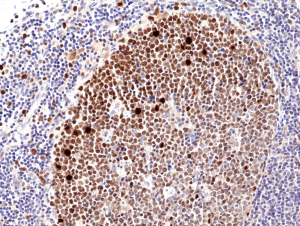
anti-KI67 (human) Rabbit Monoclonal (RM360)
| Code | Size | Price |
|---|
| REV-31-1246-00-R100 | 100 ul | £455.00 |
Quantity:
Prices exclude any Taxes / VAT
Overview
Antibody Isotype: Rabbit IgG
Antibody Clonality: Recombinant Antibody
Antibody Clone: RM360
Regulatory Status: RUO
Target Species: Human
Applications:
- Immunohistochemistry (IHC)
- Western Blot (WB)
Shipping:
Blue Ice
Storage:
+4°C
Images
Documents
Further Information
Alternate Names/Synonyms:
Proliferation Marker Protein Ki-67
Concentration:
N/A
EClass:
32160000
Form (Short):
liquid
Formulation:
Liquid. 50% Glycerol/PBS with 1% BSA and 0.09% sodium azide.
Handling Advice:
Avoid freeze/thaw cycles.
Immunogen:
A peptide corresponding to the internal region of human Ki67.
Long Description:
Recombinant Antibody. This antibody reacts to Human Antigen KI-67. Applications: WB, IHC. Source: Rabbit. Liquid. 50% Glycerol/PBS with 1% BSA and 0.09% sodium azide. Ki-67 is a nuclear protein that is expressed during various stages in the cell cycle, particularly during late G1, S, G2, and M phases. The protein has a forkhead associated domain (FHA) through which it associates with euchromatin at the perichromosomal layer, the centromeric heterochromatin and the nucleolus. Ki-67 is shown to have a cell cycle dependent topographical distribution with perinucleolar expression at G1, expression in the nuclear matrix at G2 and expression on the chromosomes during M phase. Ki-67 is commonly used as a proliferation marker because it is not detected in G0 cells, but increases steadily from G1 through mitosis. Ki-67 antibodies are useful in establishing the cell growing fraction in neoplasms. In neoplastic tissues, the prognostic value is comparable to the tritiated thymidine-labelling index. The correlation between low Ki-67 index and histologically low-grade tumors is strong. Ki-67 is routinely used as a neuronal marker of cell cycling and proliferation. In breast cancer Ki67 identifies a high proliferative subset of patients with ER-positive breast cancer who derive greater benefit from adjuvant chemotherapy.
NCBI, Uniprot Number:
P46013
Package Type:
Vial
Product Description:
Ki-67 is a nuclear protein that is expressed during various stages in the cell cycle, particularly during late G1, S, G2, and M phases. The protein has a forkhead associated domain (FHA) through which it associates with euchromatin at the perichromosomal layer, the centromeric heterochromatin and the nucleolus. Ki-67 is shown to have a cell cycle dependent topographical distribution with perinucleolar expression at G1, expression in the nuclear matrix at G2 and expression on the chromosomes during M phase. Ki-67 is commonly used as a proliferation marker because it is not detected in G0 cells, but increases steadily from G1 through mitosis. Ki-67 antibodies are useful in establishing the cell growing fraction in neoplasms. In neoplastic tissues, the prognostic value is comparable to the tritiated thymidine-labelling index. The correlation between low Ki-67 index and histologically low-grade tumors is strong. Ki-67 is routinely used as a neuronal marker of cell cycling and proliferation. In breast cancer Ki67 identifies a high proliferative subset of patients with ER-positive breast cancer who derive greater benefit from adjuvant chemotherapy.
Purity:
Protein A purified.
Source / Host:
Rabbit
Specificity:
This antibody reacts to Human Antigen KI-67.
Transportation:
Non-hazardous
UNSPSC Category:
Primary Antibodies
UNSPSC Number:
12352203
Use & Stability:
Stable for at least 1 year after receipt when stored at -20°C.


