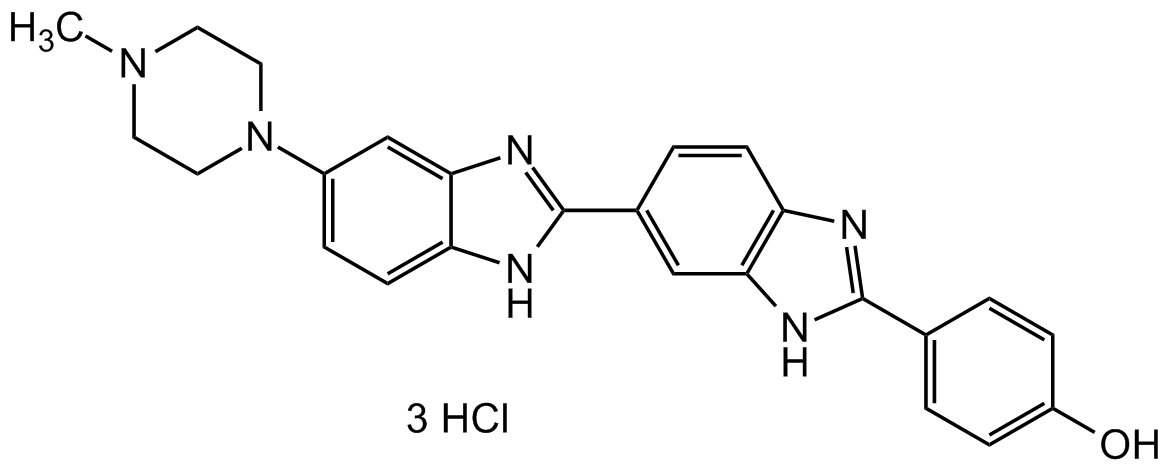
Hoechst 33258 Solution
| Code | Size | Price |
|---|
| CDX-B0446-L005 | 5 ml | £126.00 |
Quantity:
Prices exclude any Taxes / VAT
Overview
Regulatory Status: RUO
Shipping:
AMBIENT
Storage:
+4°C
Images
Documents
Further Information
Alternate Names/Synonyms:
4-[5-(4-Methyl-1-piperazinyl)-1H,1'H-2,5'-bibenzimidazol-2'-yl]phenol trihydrochloride; BisBenzimide H 33258; HOE 33258
Appearance:
Liquid.
CAS:
23491-45-4
Concentration:
1mg/ml in water
EClass:
32160000
Form (Short):
liquid
GHS Symbol:
GHS07
Handling Advice:
Protect from light and moisture.
Hazards:
H302, H315, H319
InChi:
InChI=1S/C25H24N6O/c1-30-10-12-31(13-11-30)18-5-9-21-23(15-18)29-25(27-21)17-4-8-20-22(14-17)28-24(26-20)16-2-6-19(32)7-3-16/h2-9,14-15,32H,10-13H2,1H3,(H,26,28)(H,27,29)
InChiKey:
INAAIJLSXJJHOZ-UHFFFAOYSA-N
Long Description:
Chemical. CAS: 23491-45-4. Formula: C25H24N6O . 3HCl. MW: 533.88. The Hoechst stains are a family of fluorescent stains for labeling DNA in fluorescence microscopy. The blue fluorescent Hoechst dye is a cell permeable nucleic acid stain that has multiple applications, including sensitive detection of DNA in the presence of RNA in agarose gels, automated DNA determination, sensitive determination of cell number and chromosome sorting. Useful vital stain for the flow cytometric recognition of DNA damage and other viability measurements by monitoring the emission spectral shifts of the dyes. Because this fluorescent stain labels DNA, it can also be used to visualize nuclei and mitochondria. This dye is commonly used for determining the DNA content of viable cells without detergent treatment or fixation and for fluorescence microscopy or flow cytometry. Hoechst 33258 is a cell-permeable, benzimidazole dye that binds to the minor groove of double stranded DNA with preference for adenine and thymine-rich sequences. It emits blue fluorescence (excitation 352nm/emission max 461nm) when bound to DNA in either live or fixed cells and is useful as a marker of nuclei for cell cycle studies and to distinguish nuclear morphology in apoptotic cells.
MDL:
MFCD00012679
Molecular Formula:
C25H24N6O . 3HCl
Molecular Weight:
533.88
Package Type:
Vial
Precautions:
P305 + P351 + P338
Product Description:
The Hoechst stains are a family of fluorescent stains for labeling DNA in fluorescence microscopy. The blue fluorescent Hoechst dye is a cell permeable nucleic acid stain that has multiple applications, including sensitive detection of DNA in the presence of RNA in agarose gels, automated DNA determination, sensitive determination of cell number and chromosome sorting. Useful vital stain for the flow cytometric recognition of DNA damage and other viability measurements by monitoring the emission spectral shifts of the dyes. Because this fluorescent stain labels DNA, it can also be used to visualize nuclei and mitochondria. This dye is commonly used for determining the DNA content of viable cells without detergent treatment or fixation and for fluorescence microscopy or flow cytometry. Hoechst 33258 is a cell-permeable, benzimidazole dye that binds to the minor groove of double stranded DNA with preference for adenine and thymine-rich sequences. It emits blue fluorescence (excitation 352nm/emission max 461nm) when bound to DNA in either live or fixed cells and is useful as a marker of nuclei for cell cycle studies and to distinguish nuclear morphology in apoptotic cells.
Purity:
>98% (HPLC, TLC)
Signal Word:
Warning
SMILES:
CN(CC1)CCN1C2=CC=C(NC(C3=CC(NC(C4=CC=C(O)C=C4)=N5)=C5C=C3)=N6)C6=C2
Solubility Chemicals:
Soluble in water.
Source / Host:
Synthetic
Transportation:
Non-hazardous
UNSPSC Category:
Fluorescent Reagents
UNSPSC Number:
41105331
Use & Stability:
Stable for at least 2 years after receipt when stored at +4°C.
References
(1) A. Kumar et al.: In Vitro Cell. & Developm. Biol. 44(7), 189 (2008) | (2) D. Weidmann et al.: Meth. Enzym. 446, 277 (2008) | (3) D. Plesca et al.: Meth. Enzym. 446, 107 (2008) | (4) K.H. Elstein et al.: Exp. Cell. Res. 211, 322 (1994) | (5) Y.-J. Kim et al.: Anal. Biochem. 174, 168 (1988) | (6) C. Labarca et al.: Anal. Biochem. 102, 344 (1980) | (7) S.A. Latt et al.: J. Histochem. Cytochem. 24, 24 (1976)


