
Anti-LC3 (Human) pAb

| Code | Size | Price |
|---|
| MBL-PM036 | 100 ul | £374.00 |
Quantity:
Prices exclude any Taxes / VAT
Overview
Host Type: Rabbit
Antibody Isotype: IgG
Antibody Clonality: Polyclonal
Regulatory Status: RUO
Target Species:
- Hamster
- Human
- Mouse
- Rat
Applications:
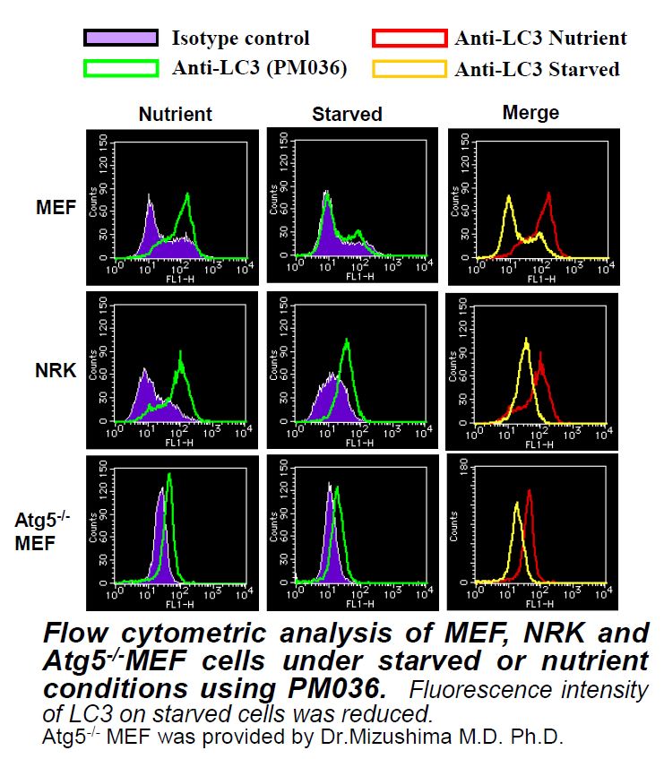
- Flow Cytometry
- Immunocytochemistry (ICC)
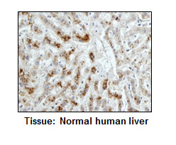
- Immunohistochemistry (IHC)
- Immunoprecipitation (IP)
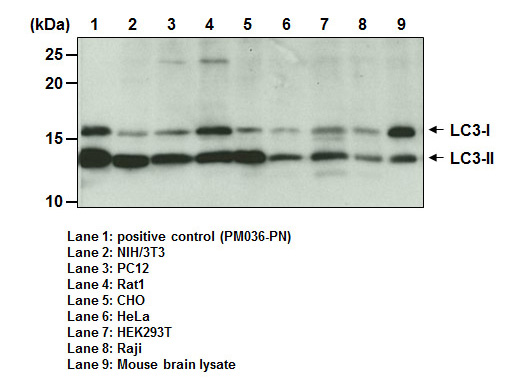
- Western Blot (WB)
Shipping:
4°C
Storage:
-20°C


Documents
Further Information
Applications:
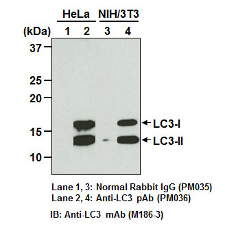
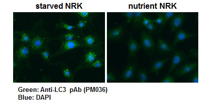
WB - 1:1000 (chemiluminescence detection system) IP - 2 uL/300 uL of cell extract from 1x10⁷ cells FCM - 1:200 (final concentration) ICC - 1:500-1:1000 IHC - 1:1000-1:2000 (heat treatment required for paraffin)
Background:
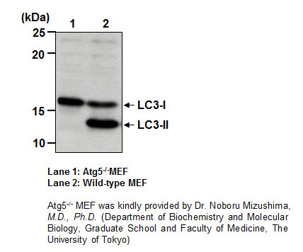
Macroautophagy mediates the bulk degradation of cytoplasmic components. These components are delivered to lysosomes via autophagosomes. The microtubule-associated protein 1 light chain 3 (LC3), a homologue of yeast Atg8 (Aut7/Apg8), localizes to autophagosomal membranes after post-translational modifications. The C-terminal fragment of LC3 is cleaved immediately following synthesis to yield a cytosolic form called LC3-I (18 kDa). A subpopulation of LC3-I is further converted to an autophagosome-associating form, LC3-II (16 kDa). This antibody can detect both forms of LC3.
Formulation:
100 ul volume of PBS containing 50% glycerol, pH 7.2. No preservative is contained.
Gene IDs:
Human: 8163184557440730 Mouse: 6744366734 Rat: 64862, 362245
Immunogen Translated:
Recombinant human LC3 (MAP1LC3B :1-120 a.a.)
Reactivity:
This antibody reacts with LC3 (MAP1LC3A, B, C) on Western blotting, Immunoprecipitation, Immunohistochemistry, Immunocytochemistry and Flow cytomrtry. It does not react with GABARAP and GATE-16.
Shelf Life:
1 year
Source:
This antibody was purified from rabbit serum using protein A agarose. The rabbit was immunized with the recombinant human LC3 [MAP1LC3B (1-120 aa)].
Target:
LC3
References
1) Tabata, K., et al., Mol. Biol. Cell 21, 4162-4172 (2010)
2) Kiyono, K., et al., Cancer Res. 69, 8844-8852 (2009)
3) Saitoh, T., et al., Nature 456, 264-268 (2008)
4) Wan, G., et al., J. Biol. Chem. 283, 21540-21549 (2008)
5) Ohneet, Y., et al., J. Biol. Chem. 283, 31861-31870 (2008)
6) Kabeya, Y., et al., J. Cell Sci. 117, 2805-2812 (2004)
7) Mizushima, N., et al., Mol. Biol. Cell 15, 1101-1111 (2004)
8) Mizushima, N., et al., J. Cell Biol. 152, 657-667 (2001)
9) Kabeya, Y., et al., EMBO J. 19, 5720-5728 (2000)