PD-1 Monoclonal Antibody
| Code | Size | Price |
|---|
| PSI-PM-5179-0.02mg | 0.02mg | £150.00 |
Quantity:
| PSI-PM-5179-0.1mg | 0.1mg | £515.00 |
Quantity:
Prices exclude any Taxes / VAT
Overview
Host Type: Mouse
Antibody Isotype: IgG1
Antibody Clonality: Monoclonal
Antibody Clone: 12A7D7
Regulatory Status: RUO
Applications:
- Enzyme-Linked Immunosorbent Assay (ELISA)
- Immunohistochemistry (IHC)
- Western Blot (WB)
Images
Documents
Further Information
Additional Names:
PD-1 Antibody [12A7D7] : PD1, PD-1, CD279, SLEB2, hPD-1, hPD-l, hSLE1
Application Note:
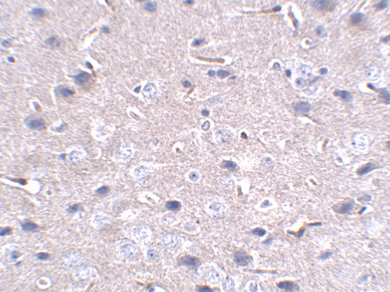
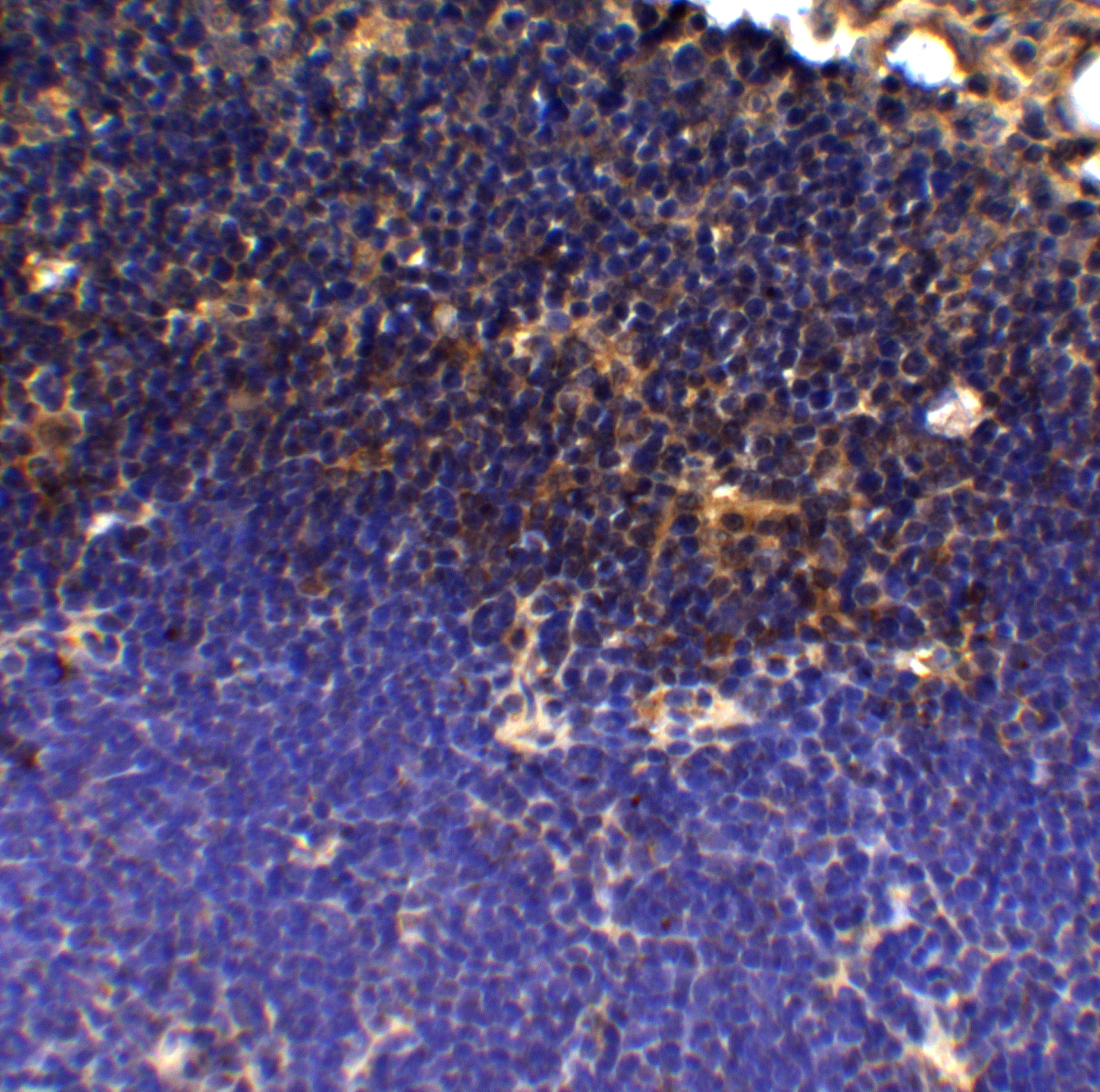
PD-1 antibody can be used for detection of PD-1 by Western blot at 1 μg/mL. Antibody can also be used for immunohistochemistry starting at 2.5 μg/mL.
Antibody validated: Western Blot in mouse samples and Immunohistochemistry in mouse samples. All other applications and species not yet tested.
Antibody validated: Western Blot in mouse samples and Immunohistochemistry in mouse samples. All other applications and species not yet tested.
Background:
PD-1 Monoclonal Antibody: Cell-mediated immune responses are initiated by T lymphocytes that are themselves stimulated by cognate peptides bound to MHC molecules on antig en-presenting cells (APC). T-cell activation is generally self-limited as activated T cells express receptors such as PD-1 (also known as PDCD-1) that mediate inhibitory signals from the APC. PD-1 can bind two different but related ligands, PDL-1 and PDL-2. Upon binding to either of these ligands, signals generated by PD-1 inhibit the activation of the immune response in the absence of "danger signals" such as LPS or other molecules associated with bacteria or other pathogens. Evidence for this is seen in PD1-null mice who exhibit hyperactivated immune systems and autoimmune diseases. Despite its predicted molecular weight, PD-1 often migrates at higher molecular weight in SDS-PAGE.
Background References:
- Holling TM, Schooten E, and van Den Elsing PJ. Function and regulation of MHC class II molecules in T-lymphocytes: of mice and men. Hum. Immunol. 2004; 65:282-90.
- Ishida Y, Agata Y, Shibahara K, et al. Induced expression of PD-1, a novel member of the immunoglobulin gene superfamily, upon programmed cell death. EMBO J. 1992; 11:3887-95.
- Zhong X, Bai C, Gao W, et al. Suppression of expression and function of negative immune regulator PD-1 by certain pattern recognition and cytokine receptor signals associated with immune system danger. Int. Immunol. 2004; 16:1181-8.
- Nishimura H, Nose M, Hiai H, et al. Development of lupus-like autoimmune diseases by the disruption of the PD-1 gene encoding an ITIM motif-carrying immunoreceptor. Immunity 1999; 11:141-51.
Buffer:
PD-1 Monoclonal Antibody is supplied in PBS containing 0.02% sodium azide.
Concentration:
1 mg/mL
Conjugate:
Unconjugated
DISCLAIMER:
Optimal dilutions/concentrations should be determined by the end user. The information provided is a guideline for product use. This product is for research use only.
Immunogen:
A ~150 amino acid recombinant protein from near the amino terminus of mouse PD-1.
NCBI Gene ID #:
5133
NCBI Official Name:
programmed cell death 1
NCBI Official Symbol:
PDCD1
NCBI Organism:
mus musculus
Physical State:
Liquid
Protein Accession #:
EDL39922
Protein GI Number:
148707975
Purification:
PD-1 Monoclonal Antibody is Protein A purified.
Research Area:
Apoptosis
Swissprot #:
Q15116
User NOte:
Optimal dilutions for each application to be determined by the researcher.


