IRAK Antibody

ProSci
Product Code: PSI-1007
Product Group: Primary Antibodies
Supplier: ProSci
CodeSizePrice
PSI-1007-0.02mg0.02mg£150.00
Quantity:
PSI-1007-0.1mg0.1mg£449.00
Quantity:
Prices exclude any Taxes / VAT

Overview

Host Type: Rabbit
Antibody Isotype: IgG
Antibody Clonality: Polyclonal
Regulatory Status: RUO
Applications:
  • Enzyme-Linked Immunosorbent Assay (ELISA)
  • Immunofluorescence (IF)
  • Immunohistochemistry (IHC)
  • Immunoprecipitation (IP)
  • Western Blot (WB)

Images

1 / 8
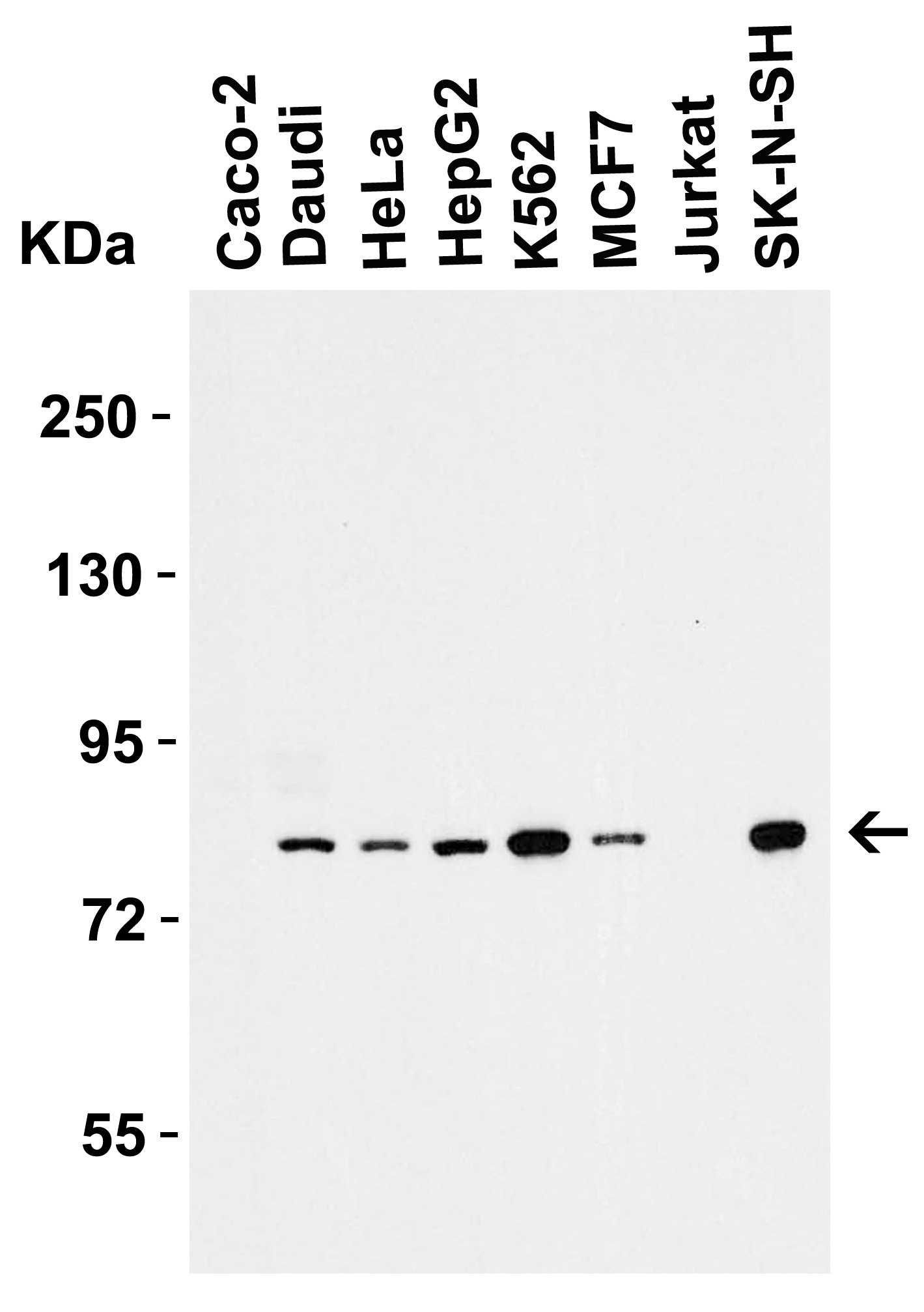
<strong>Figure 1 Western Blot Validation in Human Cell Lines</strong><br>
Loading: 15 μg of lysates per lane.
Antibodies: IRAK 1007 (1 μg/mL), 1h incubation at RT in 5% NFDM/TBST.
Secondary: Goat anti-rabbit IgG HRP conjugate at 1:10000 dilution.
2 / 8
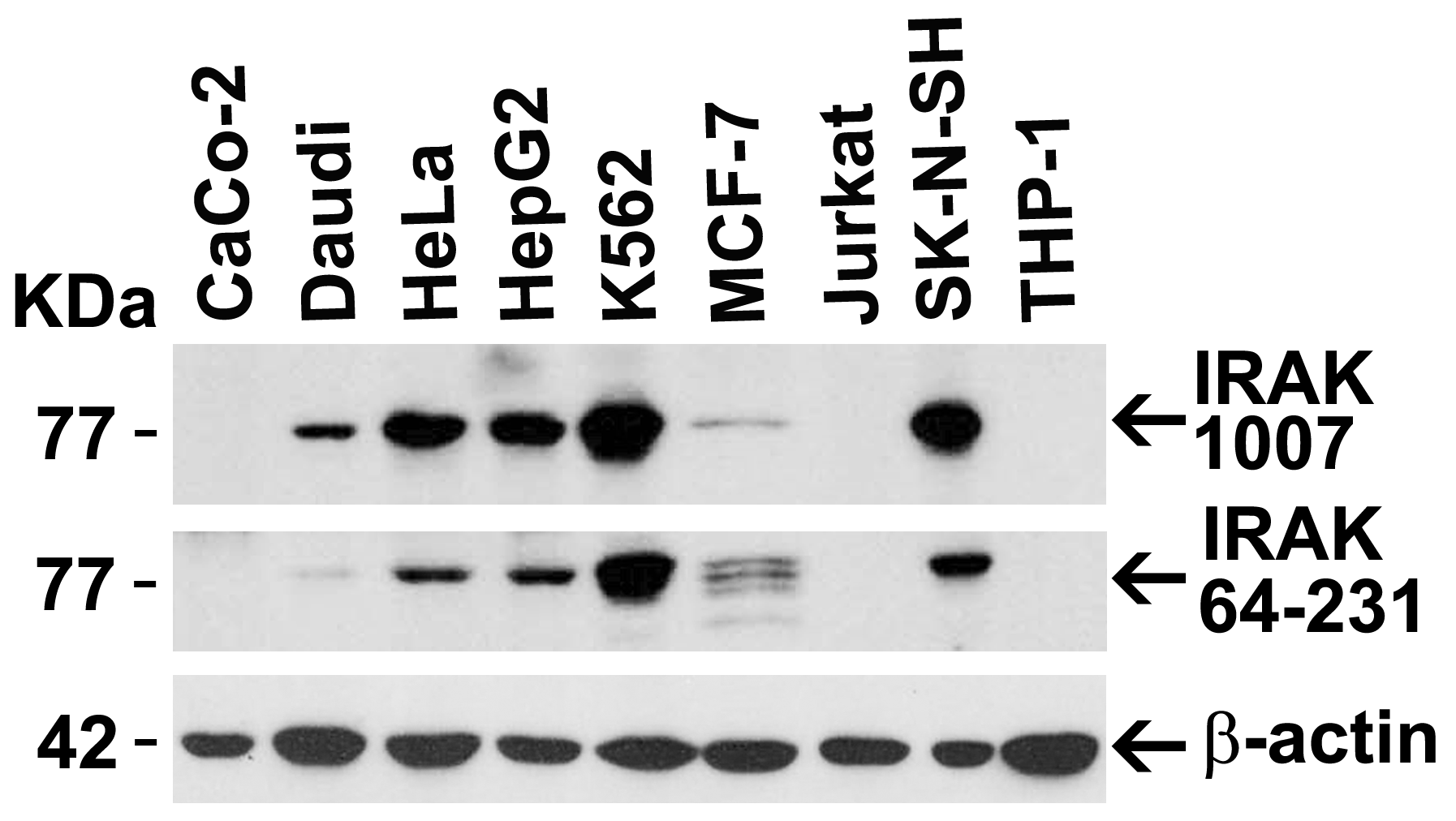
<strong>Figure 2 Independent Antibody Validation (IAV) via Protein Expression Profile in Cell Lines</strong><br>
Loading: 15 μg of lysates per lane.
Antibodies: IRAK 1007 (1 μg/mL), IRAK 64-231 (2 μg/mL), beta-actin (1 μg/mL),  1h incubation at RT  in 5% NFDM/TBST.
Secondary: Goat anti-rabbit IgG HRP conjugate at 1:10000 dilution.
3 / 8
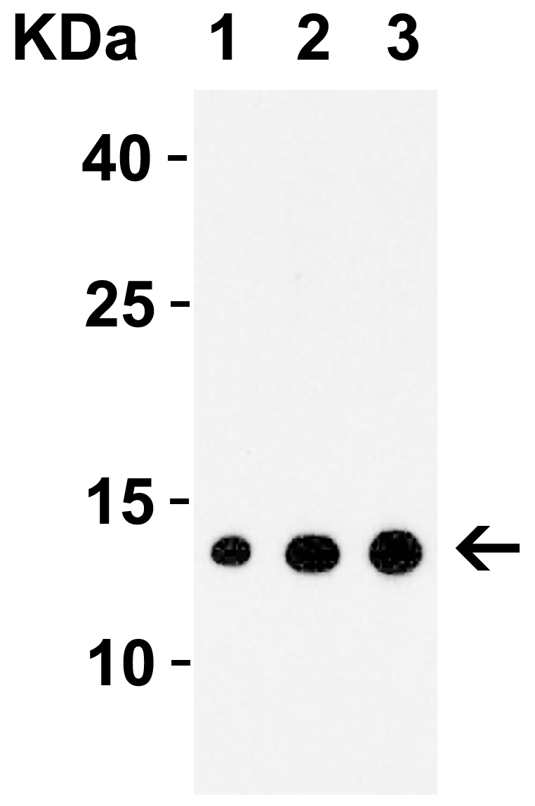
<strong>Figure 3 Western Blot Validation with Recombinant Protein</strong><br>
Loading: 30 ng of human IRAK recombinant protein per lane.
Antibodies: IRAK 1007 (1: 1 μg/mL, 2: 2 μg/mL and 3: 4 μg/mL), 1h incubation at RT  in 5% NFDM/TBST.
Secondary: Goat anti-rabbit IgG HRP conjugate at 1:10000 dilution.
4 / 8
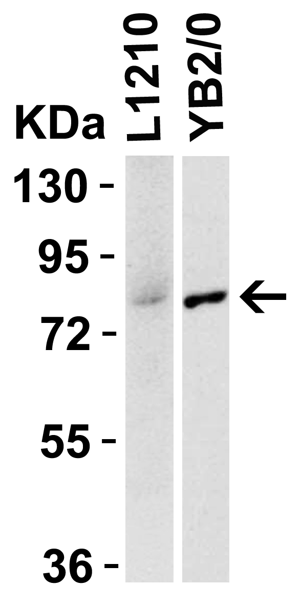
<strong>Figure 4 Species Activity in Mouse and Rat Cell Lines</strong><br>
Loading: 15 μg of lysates per lane.
Antibodies: IRAK 1007 (1 ug/mL,), 1h incubation at RT  in 5% NFDM/TBST.
Secondary: Goat anti-rabbit IgG HRP conjugate at 1:10000 dilution.
5 / 8
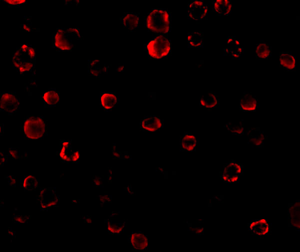
<strong>Figure 5 Immunofluorescence Validation of IRAK in Human HeLa Cells</strong><br>
Immunofluorescent analysis of 4% paraformaldehyde-fixed HeLa Cells labeling IRAK with 1007 at 20 μg/mL, followed by goat anti-rabbit IgG secondary antibody at 1/500 dilution (red).
6 / 8
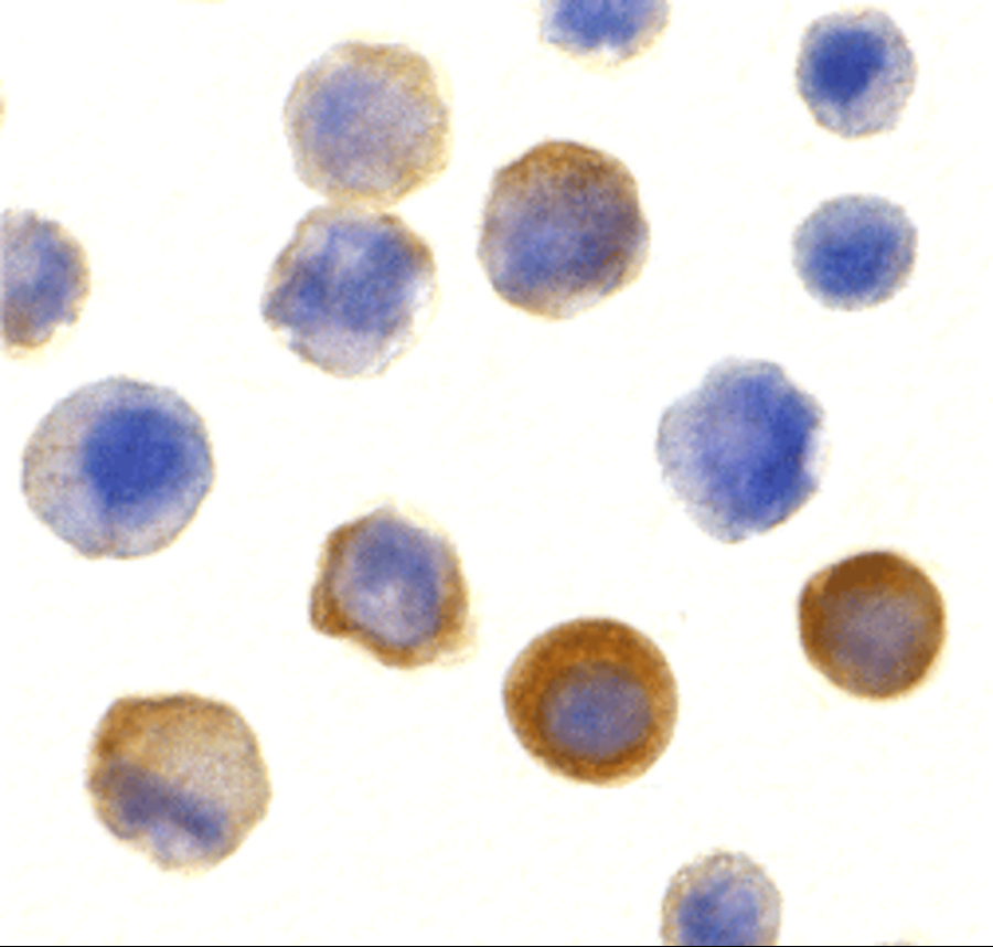
<strong>Figure 6  Immunocytochemistry Validation of IRAK in Human HeLa Cells</strong><br>
Immunocytochemical analysis of HeLa cells using anti-IRAK antibody (1007) at 10 μg/ml. Cells was fixed with formaldehyde and blocked with 10% serum for 1 h at RT; antigen retrieval was by heat mediation with a citrate buffer (pH6). Samples were incubated with primary antibody overnight at 4˚ C. A goat anti-rabbit IgG H&L (HRP) at 1/250 was used as secondary. Counter stained with Hematoxylin.
7 / 8
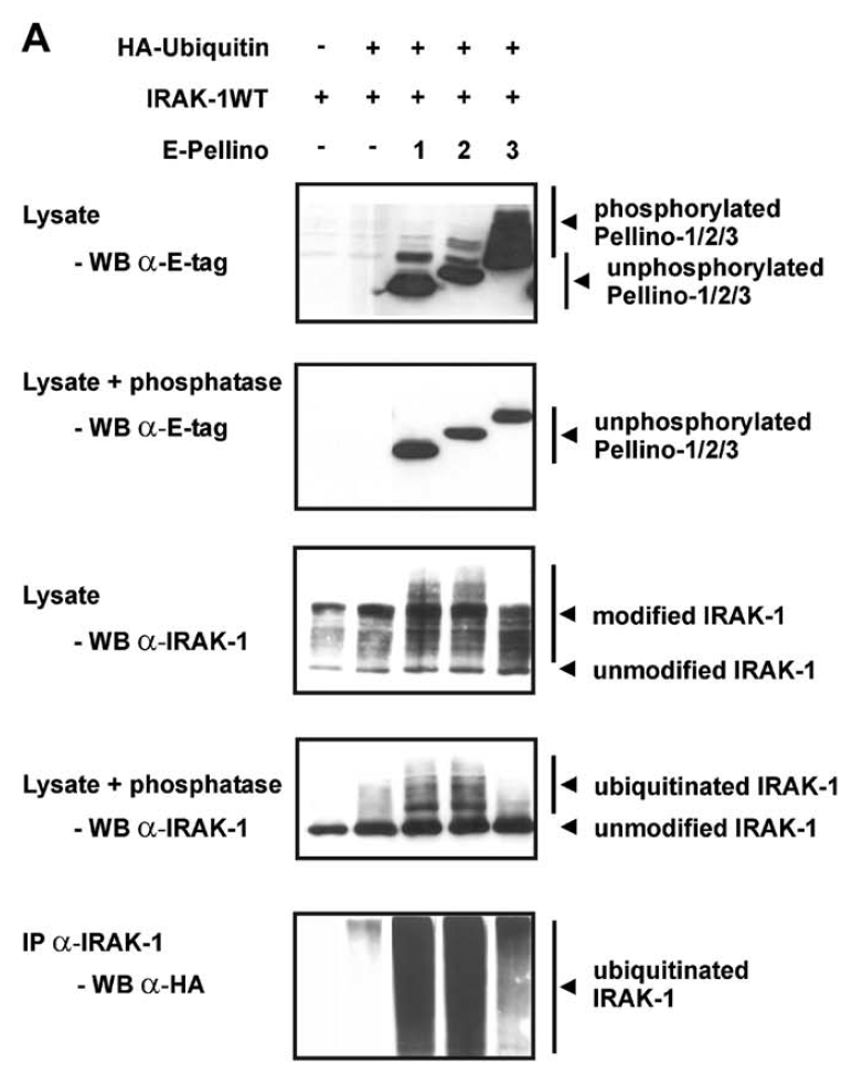
<strong>Figure 7 Immunoprecipitation and Overexpression Validation in HEK293T Cells(Schauvliege et al., 2006)</strong><br>
Co-expression of Pellino proteins and IRAK-1 leads to Pellino phosphorylation and IRAK-1 polyubiquitination. (A) E-tagged Pellino proteins were co-expressed with IRAK-1WT and HA?ubiquitin in HEK293T cells. For assessment of IRAK-1 polyubiquitination, the same cell
extracts, untreated or treated with phosphatase as described above, were analysed for slower migrating forms of IRAK-1 by Western blotting with
anti-IRAK-1 (1007). Ubiquitination was specifically detected by IRAK-1 immunoprecipitation followed by Western blotting with anti-HA antibodies.
8 / 8
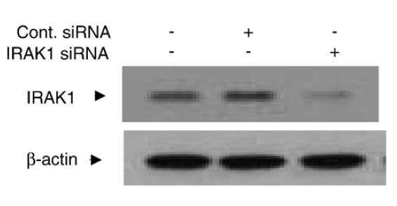
<strong>Figure 8 KD Validation in Human Chondrocytes (Ahmad et al., 2007)</strong><br>
Chondrocytes were transfected with 250 nM of IRAK1 or control siRNA for 48 h and lysates were analyzed for IRAK1 or β-actin expression levels by immunoblotting. IRAK1 signal was disrupted in IRAK1 KD lysate.

<strong>Figure 1 Western Blot Validation in Human Cell Lines</strong><br>
Loading: 15 μg of lysates per lane.
Antibodies: IRAK 1007 (1 μg/mL), 1h incubation at RT in 5% NFDM/TBST.
Secondary: Goat anti-rabbit IgG HRP conjugate at 1:10000 dilution.
<strong>Figure 2 Independent Antibody Validation (IAV) via Protein Expression Profile in Cell Lines</strong><br>
Loading: 15 μg of lysates per lane.
Antibodies: IRAK 1007 (1 μg/mL), IRAK 64-231 (2 μg/mL), beta-actin (1 μg/mL),  1h incubation at RT  in 5% NFDM/TBST.
Secondary: Goat anti-rabbit IgG HRP conjugate at 1:10000 dilution.
<strong>Figure 3 Western Blot Validation with Recombinant Protein</strong><br>
Loading: 30 ng of human IRAK recombinant protein per lane.
Antibodies: IRAK 1007 (1: 1 μg/mL, 2: 2 μg/mL and 3: 4 μg/mL), 1h incubation at RT  in 5% NFDM/TBST.
Secondary: Goat anti-rabbit IgG HRP conjugate at 1:10000 dilution.
<strong>Figure 4 Species Activity in Mouse and Rat Cell Lines</strong><br>
Loading: 15 μg of lysates per lane.
Antibodies: IRAK 1007 (1 ug/mL,), 1h incubation at RT  in 5% NFDM/TBST.
Secondary: Goat anti-rabbit IgG HRP conjugate at 1:10000 dilution.
<strong>Figure 5 Immunofluorescence Validation of IRAK in Human HeLa Cells</strong><br>
Immunofluorescent analysis of 4% paraformaldehyde-fixed HeLa Cells labeling IRAK with 1007 at 20 μg/mL, followed by goat anti-rabbit IgG secondary antibody at 1/500 dilution (red).
<strong>Figure 6  Immunocytochemistry Validation of IRAK in Human HeLa Cells</strong><br>
Immunocytochemical analysis of HeLa cells using anti-IRAK antibody (1007) at 10 μg/ml. Cells was fixed with formaldehyde and blocked with 10% serum for 1 h at RT; antigen retrieval was by heat mediation with a citrate buffer (pH6). Samples were incubated with primary antibody overnight at 4˚ C. A goat anti-rabbit IgG H&L (HRP) at 1/250 was used as secondary. Counter stained with Hematoxylin.
<strong>Figure 7 Immunoprecipitation and Overexpression Validation in HEK293T Cells(Schauvliege et al., 2006)</strong><br>
Co-expression of Pellino proteins and IRAK-1 leads to Pellino phosphorylation and IRAK-1 polyubiquitination. (A) E-tagged Pellino proteins were co-expressed with IRAK-1WT and HA?ubiquitin in HEK293T cells. For assessment of IRAK-1 polyubiquitination, the same cell
extracts, untreated or treated with phosphatase as described above, were analysed for slower migrating forms of IRAK-1 by Western blotting with
anti-IRAK-1 (1007). Ubiquitination was specifically detected by IRAK-1 immunoprecipitation followed by Western blotting with anti-HA antibodies.
<strong>Figure 8 KD Validation in Human Chondrocytes (Ahmad et al., 2007)</strong><br>
Chondrocytes were transfected with 250 nM of IRAK1 or control siRNA for 48 h and lysates were analyzed for IRAK1 or β-actin expression levels by immunoblotting. IRAK1 signal was disrupted in IRAK1 KD lysate.

Further Information

Additional Names:
IRAK Antibody: IRAK, pelle, IRAK, Interleukin-1 receptor-associated kinase 1, IRAK-1
Application Note:
WB: 1-4 μg/mL; IF: 20 μg/mL; ICC: 10 μg/mL.

Antibody validated: Western Blot in human, mouse and rat samples; Immunofluorescence and Immunocytochemistry in human samples. All other applications and species not yet tested.
Background:
IRAK Antibody: Nuclear factor kappa B (NF-κB) is a ubiquitous transcription factor and an essential mediator of gene expression during activation of immune and inflammatory responses. NF-κB mediates the expression of a great variety of genes in response to extracellular stimuli including IL-1, TNFα and LPS. A serine/threonine protein kinase associated with IL-1 receptor (IRAK) and its homologue mouse pelle-like protein kinase (mPLK) were identified recently. IRAK is associated with the IL-1 receptor subunits IL-1RI and IL-1RAcP after IL-1 binding and serves as a signaling molecule to mediate IL-1 response. IRAK mediates a signaling cascade leading to NF-κB activation by members in IL-1 family including IL-1 and a novel cytokine IL-18 (also termed IGIF).
Background References:
  • Cao et al. Science 1996;271:1128-31.
  • Trofimova et al. J Bio Chem 1996; 271: 17609-1.
  • Huang et al. Proc Natl Acad Sci USA 1997;94:12829-12832.
  • Robinson et al. Immunity 1997;7:571-581.
Buffer:
IRAK Antibody is supplied in PBS containing 0.02% sodium azide.
Concentration:
1 mg/mL
Conjugate:
Unconjugated
DISCLAIMER:
Optimal dilutions/concentrations should be determined by the end user. The information provided is a guideline for product use. This product is for research use only.
Homology:
Predicted species reactivity based on immunogen sequence: Bovine: (85%)
Immunogen:
Anti-IRAK antibody (1007) was raised against a peptide corresponding to 13 amino acids near the carboxy terminus of human IRAK.

The immunogen is located within the last 50 amino acids of IRAK.
ISOFORMS:
Human IRAK has 4 isoforms, including isoform 1 (712aa, 77kD), isoform 2 (682aa, 73kD), isoform 3 (693aa, 75kD), and isoform 4 (633aa, 68kD). Mouse IRAK has 2 isoforms, including isoform 1 (710aa, 77kD) and isoform 2 (712aa, 78kD). Rat IRAK also has one isoform (710aa, 78kD). 1007 can detect all human isoforms, mouse and rat IRAK.
NCBI Gene ID #:
3654
NCBI Official Name:
interleukin-1 receptor-associated kinase 1
NCBI Official Symbol:
IRAK1
NCBI Organism:
Homo sapiens
Physical State:
Liquid
PREDICTED MOLECULAR WEIGHT:
Predicted: 77kD

Observed: 77 kD
Protein Accession #:
P51617
Protein GI Number:
8928535
Purification:
IRAK Antibody is affinity chromatography purified via peptide column.
Research Area:
Signal Transduction,Innate Immunity
SPECIFICITY:
At least four isoforms of IRAK are known to exist; this antibody will detect all four isoforms. IRAK antibody is predicted to not cross-react with other members of the IRAK protein family.
Swissprot #:
P51617
User NOte:
Optimal dilutions for each application to be determined by the researcher.
VALIDATION:

Independent Antibody Validation in Cell lines (Figure 2) shows similar IRAK expression profile in human cell lines detected by two independent anti-IRAK antibodies that recognize different epitopes, 1007 against C-terminus domain and 64-231 against another region of C-terminus domain.  IRAK proteins are detected in the most tested cell lines at different expression levels by the two independent antibodies.

Recombinant Protein Test (Figure 3): Anti-IRAK antibodies (1007) detected human IRAK recombinant protein at different concentrations.

Immunoprecipitation validation (Figure 6): IRAK1 protein and pellino protein were immunoprecipitated by anti-IRAK antibodies (1007) in HEK293T cells with co-expression of Pellino proteins and IRAK-1.

Overexpression validation (Figure 7): IRAK1 overexpression in 293T cells was detected by anit-IRAK antibodies (1007).

KD validation (Figure 8): Anti-IRAK antibody (1007) specificity was further verified by IRAK specific knockdown. IRAK signal in human chondrocytes transfected with IRAK siRNAs was disrupted in comparison with that in cells transfected with control siRNAs.

References

  1. Schauvliege et al. Pellino proteins are more than scaffold proteins in TLR/IL-1R signalling: a role as novel RING E3-ubiquitin-ligases. FEBS Lett. 2006;580(19):4697-702. PMID: 16884718
  2. Ahmad et al. MyD88, IRAK1 and TRAF6 knockdown in human chondrocytes inhibits interleukin-1-induced matrix metalloproteinase-13 gene expression and promoter activity by impairing MAP kinase activation. Cell Signal. 2007 ;19(12):2549-57. PMID: 17905570
  3. Ahmad et al. Elevated expression of the toll like receptors 2 and 4 in obese individuals: its significance for obesity-induced inflammation. J Inflamm (Lond). 2012;9(1):48. PMID: 23191980

Related Products

Product NameProduct CodeSupplier 
IRAK PeptidePSI-1007PProSci Summary Details