BACE Antibody

ProSci
Product Code: PSI-2253
Product Group: Primary Antibodies
Supplier: ProSci
CodeSizePrice
PSI-2253-0.02mg0.02mg£150.00
Quantity:
PSI-2253-0.1mg0.1mg£449.00
Quantity:
Prices exclude any Taxes / VAT

Overview

Host Type: Rabbit
Antibody Isotype: IgG
Antibody Clonality: Polyclonal
Regulatory Status: RUO
Applications:
  • Enzyme-Linked Immunosorbent Assay (ELISA)
  • Immunohistochemistry (IHC)
  • Western Blot (WB)

Images

1 / 10
<strong>Figure 1 Western Blot Validation of BACE</strong><br>
Loading: 15 ug of lysates per lane.
Antibodies: BACE (1 μg/mL), 1h incubation at RT in 5% NFDM/TBST.
Secondary: Goat anti-rabbit IgG HRP conjugate at 1:10000 dilution.
Lane A-C: human brain tissue lysate in the absence (A) or presence (B) of blocking peptide and mouse 3T3/NIH cell lysate (C).
2 / 10
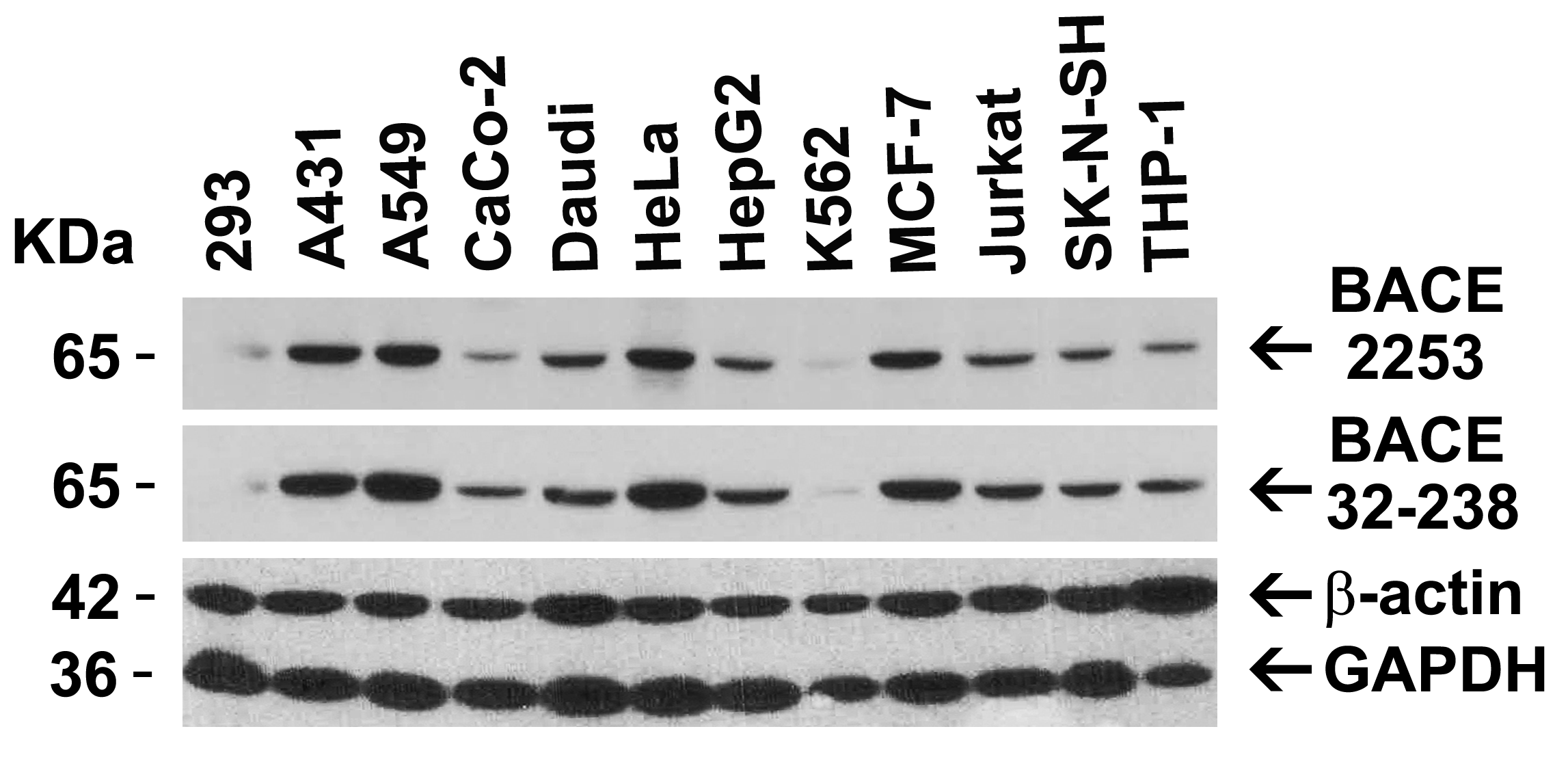
<strong>Figure 2 Independent Antibody Validation (IAV) via Protein Expression Profile in Cell Lines</strong><br>
Loading: 15 μg of lysates per lane.
Antibodies: BACE 2253 (1 μg/mL), BACE 32-238 (1 μg/mL), beta-actin (1 μg/mL), and GAPDH (0.02 μg/mL), 1h incubation at RT in 5% NFDM/TBST.
Secondary: Goat anti-rabbit IgG HRP conjugate at 1:10000 dilution.
3 / 10
<strong>Figure 3 Immunohistochemistry Validation of BACE in Mouse Brain</strong><br>
Immunohistochemical analysis of paraffin-embedded mouse brain tissue using anti-BACE antibody (2253) at 2.5 μg/ml. Tissue was fixed with formaldehyde and blocked with 10% serum for 1 h at RT; antigen retrieval was by heat mediation with a citrate buffer (pH6). Samples were incubated with primary antibody overnight at 4˚C. A goat anti-rabbit IgG H&L (HRP) at 1/250 was used as secondary. Counter stained with Hematoxylin.
4 / 10
<strong>Figure 4 Immunofluorescence Validation of BACE in 3T3/NIH Cells</strong><br>
Immunofluorescent analysis of 4% paraformaldehyde-fixed mouse 3T3/NIH cells labeling BACE with 2253 at 20 μg/mL, followed by goat anti-rabbit IgG secondary antibody at 1/500 dilution (green) and DAPI staining (blue). Image showing both membrane and cytosol staining on 3T3/NIH cells.
5 / 10
<strong>Figure 5 Immunocytochemistry Validation of BACE in 3T3/NIH Cells</strong><br>
Immunocytochemical analysis of 3T3/NIH cells using anti-BACE antibody (2253) at 10 μg/ml. Cells was fixed with formaldehyde and blocked with 10% serum for 1 h at RT; antigen retrieval was by heat mediation with a citrate buffer (pH6). Samples were incubated with primary antibody overnight at 4˚C. A goat anti-rabbit IgG H&L (HRP) at 1/250 was used as secondary. Counter stained with Hematoxylin.
6 / 10
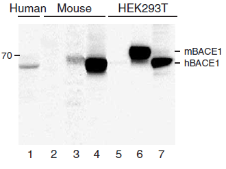
<strong>Figure 6 KO and Overexpression Validation of BACE in Human and Mouse Brain and 293 Cells. (Singer et al., 2005) </strong><br>
Western blot analysis of the BACE1 (2253) antibody's ability to recognize human and murine BACE1. The BACE1 antibody recognized both the mouse and human forms of BACE1. Lanes 1?4 are frontal cortex homogenates from human and mouse brains. Lane 1 is from a neurologically unimpaired aged human control case, lane 2 from a BACE1-deficient mouse, lane 3 from a nontransgenic mouse and lane 4 from hBACE1 transgenic mouse. Lanes 5?7 are lysates from HEK293T cells transfected with a plasmid vector expressing eGFP, mBACE1 and hBACE1, respectively.
7 / 10
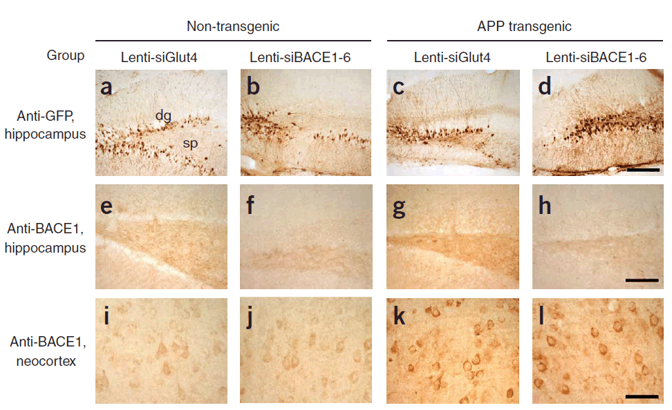
<strong>Figure 7 KD Validation of BACE in Mouse Brain (Singer et al., 2005)</strong><br> 
Characterization of the effects of lenti-siBACE1-6 expression in the brains of APP transgenic mice. (a?d) Anti-eGFP immunoreactivity in the hippocampus (the injection site) shows comparable and consistent expression of lenti-siRNA constructs in the dentate gyrus (dg) and stratus polymorphus (sp). (e) Anti-BACE1 immunoreactivity in the hippocampus of nontransgenic mice treated with lenti-siGlut4. (f) Reduced BACE1 immunostaining in the hippocampus of nontransgenic mice treated with lenti-siBACE1-6 vector. (g) Intense BACE1 immunoreactivity in the hippocampus of APP transgenic mice treated with lenti-siGlut4. (h) Reduced BACE1 expression in APP transgenic mice treated with lenti-siBACE1-6 vector. (i,j) Anti-BACE1 reacted with pyramidal cell bodies in the neocortex, which was not injected,
8 / 10
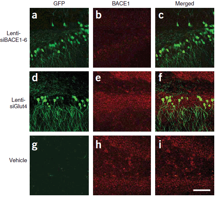
<strong>Figure 8 KD Validation of BACE in Mouse Brain (Singer et al., 2005)</strong><br> 
Immunolabeling patterns of BACE1 expression and the lenti-siRNA distribution. Sections from APP transgenic mice treated with the eGFPtagged lenti siRNA vectors (green) were co-immunolabeled with an antibody against BACE1 (red) and imaged with the LSCM. All sections are from the hippocampus of treated mice. (a?c) Lenti-siBACE1-6?treated mice. Areas within the hippocampus expressing the eGFP tagged vector have reduced BACE1 immunolabeling. (d?f) Mice treated with the eGFP-tagged control lenti-siGlut4 show unchanged expression of BACE1 in the hippocampus. (g?i) Mice treated with a saline vehicle show unchanged expression of BACE1 in the hippocampus..
9 / 10
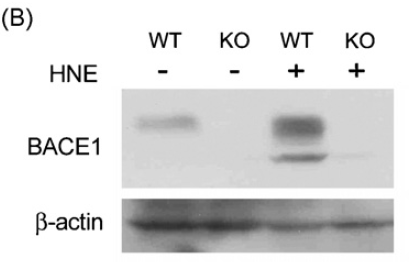
<strong>Figure 9 KO Validation of BACE in MEF Cells (Jo et al., 2010) </strong><br> 
Wildtype and BACE -/- MEFs were exposed to HNE (15_M) for 2 h. BACE1 levels were examined by Western blot with anti-BACE antibodies (2253).
10 / 10
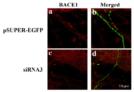
<strong>Figure 10 KD Validation of BACE in DRG (Hyun, 2007) </strong><br> 
Decreased BACE1 expression in DRG following siRNA3 transfection. DRG neurons were transfected with 1 ug siRNA3 plasmid and incubated for 48 hours in 37°˚C. DRG neurons were stained for BACE1 us?ing the Anti-BACE antibody (ProSci). (a,b) Neurons transfected with the control plas?mid pSUPER-EGFP (green) did not display any changes in BACE1 expression (red). (c,d) DRG neurons transfected with siR?NA3 displayed reduced BACE1 expression in the axon.

<strong>Figure 1 Western Blot Validation of BACE</strong><br>
Loading: 15 ug of lysates per lane.
Antibodies: BACE (1 μg/mL), 1h incubation at RT in 5% NFDM/TBST.
Secondary: Goat anti-rabbit IgG HRP conjugate at 1:10000 dilution.
Lane A-C: human brain tissue lysate in the absence (A) or presence (B) of blocking peptide and mouse 3T3/NIH cell lysate (C).
<strong>Figure 2 Independent Antibody Validation (IAV) via Protein Expression Profile in Cell Lines</strong><br>
Loading: 15 μg of lysates per lane.
Antibodies: BACE 2253 (1 μg/mL), BACE 32-238 (1 μg/mL), beta-actin (1 μg/mL), and GAPDH (0.02 μg/mL), 1h incubation at RT in 5% NFDM/TBST.
Secondary: Goat anti-rabbit IgG HRP conjugate at 1:10000 dilution.
<strong>Figure 3 Immunohistochemistry Validation of BACE in Mouse Brain</strong><br>
Immunohistochemical analysis of paraffin-embedded mouse brain tissue using anti-BACE antibody (2253) at 2.5 μg/ml. Tissue was fixed with formaldehyde and blocked with 10% serum for 1 h at RT; antigen retrieval was by heat mediation with a citrate buffer (pH6). Samples were incubated with primary antibody overnight at 4˚C. A goat anti-rabbit IgG H&L (HRP) at 1/250 was used as secondary. Counter stained with Hematoxylin.
<strong>Figure 4 Immunofluorescence Validation of BACE in 3T3/NIH Cells</strong><br>
Immunofluorescent analysis of 4% paraformaldehyde-fixed mouse 3T3/NIH cells labeling BACE with 2253 at 20 μg/mL, followed by goat anti-rabbit IgG secondary antibody at 1/500 dilution (green) and DAPI staining (blue). Image showing both membrane and cytosol staining on 3T3/NIH cells.
<strong>Figure 5 Immunocytochemistry Validation of BACE in 3T3/NIH Cells</strong><br>
Immunocytochemical analysis of 3T3/NIH cells using anti-BACE antibody (2253) at 10 μg/ml. Cells was fixed with formaldehyde and blocked with 10% serum for 1 h at RT; antigen retrieval was by heat mediation with a citrate buffer (pH6). Samples were incubated with primary antibody overnight at 4˚C. A goat anti-rabbit IgG H&L (HRP) at 1/250 was used as secondary. Counter stained with Hematoxylin.
<strong>Figure 6 KO and Overexpression Validation of BACE in Human and Mouse Brain and 293 Cells. (Singer et al., 2005) </strong><br>
Western blot analysis of the BACE1 (2253) antibody's ability to recognize human and murine BACE1. The BACE1 antibody recognized both the mouse and human forms of BACE1. Lanes 1?4 are frontal cortex homogenates from human and mouse brains. Lane 1 is from a neurologically unimpaired aged human control case, lane 2 from a BACE1-deficient mouse, lane 3 from a nontransgenic mouse and lane 4 from hBACE1 transgenic mouse. Lanes 5?7 are lysates from HEK293T cells transfected with a plasmid vector expressing eGFP, mBACE1 and hBACE1, respectively.
<strong>Figure 7 KD Validation of BACE in Mouse Brain (Singer et al., 2005)</strong><br> 
Characterization of the effects of lenti-siBACE1-6 expression in the brains of APP transgenic mice. (a?d) Anti-eGFP immunoreactivity in the hippocampus (the injection site) shows comparable and consistent expression of lenti-siRNA constructs in the dentate gyrus (dg) and stratus polymorphus (sp). (e) Anti-BACE1 immunoreactivity in the hippocampus of nontransgenic mice treated with lenti-siGlut4. (f) Reduced BACE1 immunostaining in the hippocampus of nontransgenic mice treated with lenti-siBACE1-6 vector. (g) Intense BACE1 immunoreactivity in the hippocampus of APP transgenic mice treated with lenti-siGlut4. (h) Reduced BACE1 expression in APP transgenic mice treated with lenti-siBACE1-6 vector. (i,j) Anti-BACE1 reacted with pyramidal cell bodies in the neocortex, which was not injected,
<strong>Figure 8 KD Validation of BACE in Mouse Brain (Singer et al., 2005)</strong><br> 
Immunolabeling patterns of BACE1 expression and the lenti-siRNA distribution. Sections from APP transgenic mice treated with the eGFPtagged lenti siRNA vectors (green) were co-immunolabeled with an antibody against BACE1 (red) and imaged with the LSCM. All sections are from the hippocampus of treated mice. (a?c) Lenti-siBACE1-6?treated mice. Areas within the hippocampus expressing the eGFP tagged vector have reduced BACE1 immunolabeling. (d?f) Mice treated with the eGFP-tagged control lenti-siGlut4 show unchanged expression of BACE1 in the hippocampus. (g?i) Mice treated with a saline vehicle show unchanged expression of BACE1 in the hippocampus..
<strong>Figure 9 KO Validation of BACE in MEF Cells (Jo et al., 2010) </strong><br> 
Wildtype and BACE -/- MEFs were exposed to HNE (15_M) for 2 h. BACE1 levels were examined by Western blot with anti-BACE antibodies (2253).
<strong>Figure 10 KD Validation of BACE in DRG (Hyun, 2007) </strong><br> 
Decreased BACE1 expression in DRG following siRNA3 transfection. DRG neurons were transfected with 1 ug siRNA3 plasmid and incubated for 48 hours in 37°˚C. DRG neurons were stained for BACE1 us?ing the Anti-BACE antibody (ProSci). (a,b) Neurons transfected with the control plas?mid pSUPER-EGFP (green) did not display any changes in BACE1 expression (red). (c,d) DRG neurons transfected with siR?NA3 displayed reduced BACE1 expression in the axon.

Further Information

Additional Names:
BACE Antibody: ASP2, BACE, HSPC104, KIAA1149, Beta-secretase 1, Aspartyl protease 2, ASP2
Application Note:
WB: 1 μg/mL; IHC-P: 2.5 μg/mL; ICC: 10 μg/mL; IF: 20 μg/mL.

Antibody validated: Western Blot in human and mouse samples; Immunohistochemistry, Immunocytochemistry and Immunofluorescence in mouse samples. All other applications and species not yet tested.
Background:
BACE Antibody: Accumulation of the amyloid-beta (Abeta) plaque in the cerebral cortex is a critical event in the pathogenesis of Alzheimer's disease. Abeta peptide is generated by proteolytic cleavage of the beta-amyloid protein precursor (APP) at beta- and gamma-sites by two proteases. APP is first cleaved by beta-secretase, producing a soluble derivative of the protein and a membrane anchored 99-amino acid carboxy-terminal fragment (C99). The C99 fragment serves as substrate for gamma-secretase to generate the 4 kDa amyloid-beta peptide, which is deposited in the brains of all suffers of Alzheimer's disease. The long-sought beta-secretase was recently identified by several groups independently and designated beta-site APP cleaving enzyme (BACE) and aspartyl protease 2 (Asp2). BACE/Asp2 is a novel transmembrane aspartic protease and colocalizes with APP.
Background References:
  • Vassar et al. Science 1999;286:735-41
  • Hussain et al. Mol Cell Neurosci 1999;14:419-27
  • Yan et al. Nature 1999;402:533-7
  • Sinha et al. Nature 1999;402:537-40
Buffer:
BACE Antibody is supplied in PBS containing 0.02% sodium azide.
Concentration:
1 mg/mL
Conjugate:
Unconjugated
DISCLAIMER:
Optimal dilutions/concentrations should be determined by the end user. The information provided is a guideline for product use. This product is for research use only.
Homology:
Predicted species reactivity based on immunogen sequence: Guinea pig: (94%), Rat: (94%), Bovine: (94%)
Immunogen:
Anti-BACE antibody (2253) was raised against a peptide corresponding to 17 amino acids near the carboxy terminus of human BACE.

The immunogen is located within the last 50 amino acids of BACE.
ISOFORMS:
Human BACE has 6 isoforms, including isoform A (501aa, 55.8kD), isoform B (476aa, 52.9kD), isoform C (457aa, 51.1kD), isoform D (432aa, 48.2kD), isoform 5 (401aa, 45kD) and isoform 6 (376aa, 42.2kD). This antibody detects all human isoforms. Mouse BACE has only 1 isoform (501aa, 55.7kD). Rat BACE has only one isoform identified so far (501aa, 55.8kD).
NCBI Gene ID #:
23621
NCBI Official Name:
beta-site APP-cleaving enzyme 1
NCBI Official Symbol:
BACE1
NCBI Organism:
Homo sapiens
Physical State:
Liquid
PREDICTED MOLECULAR WEIGHT:
Predicted: 55kD

Observed: 65kD (Post-modification: 4 N-linked glycosylation)
Protein Accession #:
AF190725
Protein GI Number:
6118538
Purification:
BACE Antibody is affinity chromatography purified via peptide column.
Research Area:
Neuroscience,Cancer
Swissprot #:
P56817
User NOte:
Optimal dilutions for each application to be determined by the researcher.
VALIDATION:

Independent Antibody Validation in Cell lines (Figure 2) shows similar BACE expression profile in human cell lines detected by two independent anti-BACE antibodies that recognize different epitopes, 2253 against C-terminus domain and  32-238 against recombinant fragment protein.  BACE proteins are detected in all the tested cell lines except K562 at different expression levels by the two independent antibodies.  

KO validation (Figure 6,9): Anti-BACE antibody (2253) specificity was further verified by BACE KO mice (figure6) and KO cell line (figure9). BACE signal was not detected in BACE KO mice and KO cell line.

KD validation (Figure 7,8,10): Anti-BACE antibody (2253) specificity was verified by BACE specific siRNA knockdown. BACE signal in mouse brain injected with BACE siRNAs and DRG transfected with BACE siRNAs was disrupted in comparison with control.

Overexpression validation (Figure 6): Anti-BACE antibody (2253) detected high expression levels of BACE in 293 cells transfected with hBACE or mBACE (figure6) as compared to eGFP transfected cells.

References

  1. Singer et al. Targeting BACE1 with siRNAs ameliorates Alzheimer disease neuropathology in a transgenic model. Nat Neurosci. 2005;8(10):1343-9. PMID: 16136043
  2. Jo et al.Evidence that gamma-secretase mediates oxidative stress-induced beta-secretase expression in Alzheimer's disease. Neurobiol Aging. 2010;31(6):917-25. PMID: 18687504
  3. Hyun. RNA Interference Against BACE1 Suppresses BACE1 and A? Expression in PC12 Cells and DRG Neurons. Stanford Journal of Neuroscience. 2007; 1(1):2-8.PMID:
  4. Okada et al. Proteomic identification of sorting nexin 6 as a negative regulator of BACE1-mediated APP processing. FASEB J. 2010;24(8):2783-94PMID: 20354142
  5. Liu et al. Glu11 site cleavage and N-terminally truncated A beta production upon BACE overexpression. Biochemistry. 2002;41(9):3128-36PMID: 11863452
  6. Colombo et al. JNK regulates APP cleavage and degradation in a model of Alzheimer's disease. Neurobiol Dis. 2009;33(3):518-25PMID: 19166938
  7. Bettegazzi et al. ?-Secretase activity in rat astrocytes:translational block of BACE1 and modulation of BACE2 expression. Eur J Neurosci. 2011;33(2):236-43.PMID: 21073551
  8. Lichtenthaler et al. The cell adhesion protein P-selectin glycoprotein ligand-1 is a substrate for the aspartyl protease BACE1. J Biol Chem. 2003;278(49):48713-9PMID: 14507929
  9. Lee et al. Secretion and intracellular generation of truncated Abeta in beta site amyloid-beta precursor protein-cleaving enzyme expressing human neurons. J Biol Chem. 2003;278(7):4458-66PMID: 12480937
  10. Rockenstein et al. High beta-secretase activity elicits neurodegeneration in transgenic mice despite reductions in amyloid-beta levels: implications for the treatment of Alzheimer disease. J Biol Chem. 2005;280(38):32957-67. PMID: 16027115
  11. Catania et al. The amyloidogenic potential and behavioral correlates of stress. Mol Psychiatry. 2009;14(1):95-105PMID: 17912249
  12. Parisiadou et al.Homer2 and Homer3 interact with amyloid precursor protein and inhibit Abeta production. Neurobiol Dis. 2008;30(3):353-64PMID: 18387811
  13. Knock et al. SUMO1 impact on Alzheimer disease pathology in an amyloid depositing mouse model. Neurobiol Dis. 2018;110:154-165PMID: 29217476
  14. Debatin et al. Association between deposition of beta-amyloid and pathological prion protein in sporadic Creutzfeldt-Jakob disease. Neurodegener Dis. 2008;5(6):347-54.PMID: 18349519
  15. Gwon etal. Selenium attenuates A beta production and A beta-induced neuronal death. Neurosci Lett. 2010;469(3):391-5. PMID: 20026385

Related Products

Product NameProduct CodeSupplier 
BACE Peptide PSI-2253PProSci Summary Details