PUMA Antibody
| Code | Size | Price |
|---|
| PSI-3041-0.02mg | 0.02mg | £150.00 |
Quantity:
| PSI-3041-0.1mg | 0.1mg | £449.00 |
Quantity:
Prices exclude any Taxes / VAT
Overview
Host Type: Rabbit
Antibody Isotype: IgG
Antibody Clonality: Polyclonal
Regulatory Status: RUO
Applications:
- Enzyme-Linked Immunosorbent Assay (ELISA)
- Immunofluorescence (IF)
- Immunohistochemistry (IHC)
- Western Blot (WB)
Images
Documents
Further Information
Additional Names:
PUMA Antibody: JFY1, PUMA, JFY-1, Bcl-2-binding component 3
Application Note:
WB: 1-4 μg/mL; IF: 2 μg/mL; ICC: 1 μg/mL.
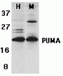
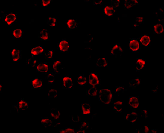
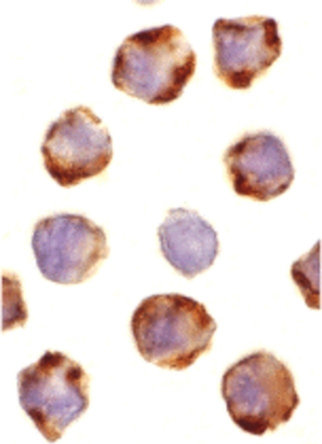
Antibody validated: Western Blot in human and mouse samples; Immunocytochemistry and Immunofluorescence in human samples. All other applications and species not yet tested.
Antibody validated: Western Blot in human and mouse samples; Immunocytochemistry and Immunofluorescence in human samples. All other applications and species not yet tested.
Background:
PUMA Antibody: Apoptosis is related to many diseases and development. The p53 tumor-suppressor protein induces apoptosis through transcriptional activation of several genes. A novel p53 inducible pro-apoptotic gene was identified recently and designated PUMA (for p53 upregulated modulator of apoptosis) and bbc3 (for Bcl-2 binding component 3) in human and mouse. PUMA/bbc3 is one of the pro-apoptotic Bcl-2 family members including Bax and Noxa, which are also transcriptional targets of p53 (1). The PUMA gene encodes two BH3 domain-containing proteins termed PUMA-alpha and PUMA-beta (2). PUMA proteins bind Bcl-2, localize to the mitochondria, and induce cytochrome c release and apoptosis in response to p53. PUMA may be a direct mediator of p53-induced apoptosis.
Background References:
- Nakano and Vousden. Mol Cell. 2001; 7(3)683-94
- Han et al. Proc Natl Acad Sci U S A. 2001;98(20):11318-23.
Buffer:
PUMA Antibody is supplied in PBS containing 0.02% sodium azide.
Concentration:
1 mg/mL
Conjugate:
Unconjugated
DISCLAIMER:
Optimal dilutions/concentrations should be determined by the end user. The information provided is a guideline for product use. This product is for research use only.
Homology:
Predicted species reactivity based on immunogen sequence: Rat: (100%)
Immunogen:
Anti-PUMA antibody (3041) was raised against a peptide corresponding to 14 amino acids near the carboxyl terminus human PUMA isoform 1.
The immunogen is located within the last 50 amino acids of PUMA.
The immunogen is located within the last 50 amino acids of PUMA.
ISOFORMS:
Human PUMA has 4 isoforms, including isoform 1 (193aa, 21kD), isoform 2 (131aa, 14kD), isoform 3 (101aa, 10kD) and isofrom 4 (261aa, 27kD). This antibody detects human isoform 1&2, mouse and rat PUMA (193aa, 21kD for both of them).
NCBI Gene ID #:
27113
NCBI Official Name:
BCL2 binding component 3
NCBI Official Symbol:
BBC3
NCBI Organism:
Homo sapiens
Physical State:
Liquid
PREDICTED MOLECULAR WEIGHT:
23 kDa
Protein Accession #:
NP_055232
Protein GI Number:
15193488
Purification:
PUMA Antibody is affinity chromatography purified via peptide column.
Research Area:
Apoptosis,Neuroscience,Autophagy,Cancer
SPECIFICITY:
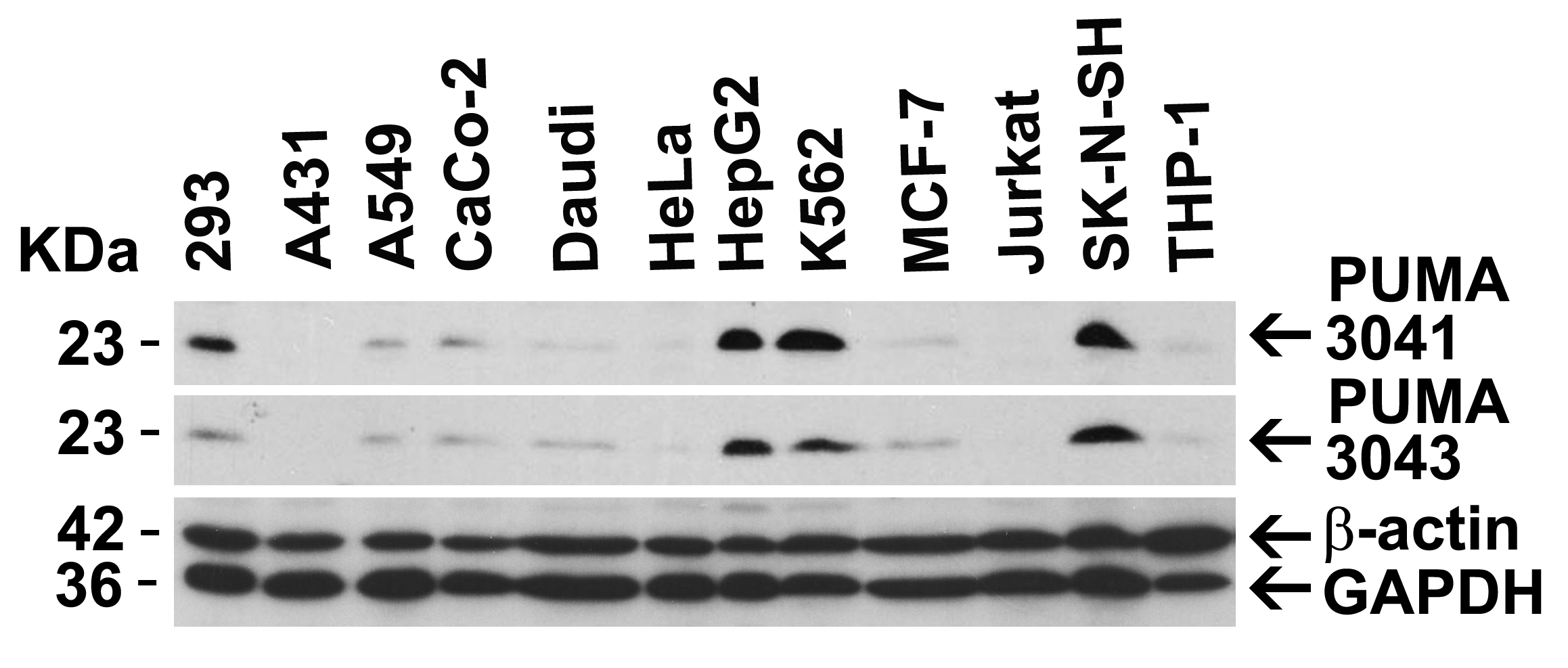
A lower band at approximately 16 kDa was detected in Daudi and K562 cells, which may represent the PUMA isoform 2.
Swissprot #:
Q96PG8
User NOte:
Optimal dilutions for each application to be determined by the researcher.
VALIDATION:
Independent Antibody Validation (Figure 2) shows similar PUMA expression profile in both human and mouse cell lines detected by two independent anti-PUMA antibodies that recognize different epitopes, 3041 against C-terminus and 3043 against the N-terminus. PUMA proteins are detected in most of the cell lines with different expression levels by the two independent antibodies.
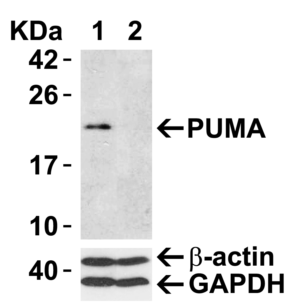
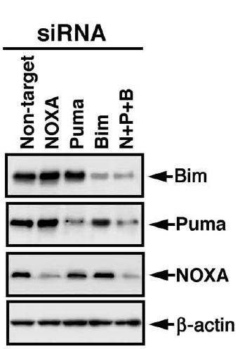
siRNA Knockdown Validation (Figure 3): Anti-PUMA antibody (3041) specificity was further verified by PUMA specific siRNA knockdown. PUMA signal in HeLa cells transfected with PUMA siRNAs was much weaker in comparison with that in HeLa cells transfected with control siRNAs.
References
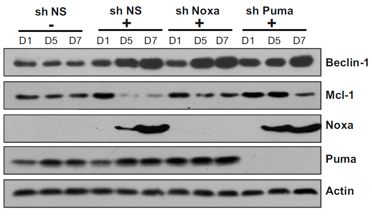
- Elgendy et al. Oncogenic Ras-induced expression of Noxa and beclin-1 promotes autophagic cell death and limits clonogenic survival. Mol. Cell. 2011;42(1):23-35. PMID: 21353614
- Han et al. Regulation of mitochondrial apoptotic events by p53-mediated disruption of complexes between anti-apoptotic Bcl-2 members and Bim. J.B.C. 2010; 285(29):22473-83. PMID: 20404322
- Sabirzhanov et al. Downregulation of miR-23a and miR-27a following experimental traumatic brain injury induces neuronal cell death through activation of proapoptotic Bcl-2 proteins. J Neurosci. 2014;34(30):10055-71. PMID: 25057207
- O'Neill et al. Inactivation of prosurvival Bcl-2 proteins activates Bax/Bak through the outer mitochondrial membrane. Genes Dev. 2016;30(8):973-88. PMID: 27056669
- Sabirzhanov et al. miR-711 upregulation induces neuronal cell death after traumatic brain injury. Cell Death Differ. 2016;23(4):654-68. PMID: 26470728
- Shi et al. NSC-87877 inhibits DUSP26 function in neuroblastoma resulting in p53-mediated apoptosis. Cell Death Dis. 2015;6:e1841. PMID: 26247726
- Meng et al. Dose-response transition from cell cycle arrest to apoptosis with selective degradation of Mdm2 and p21WAF1/CIP1 in response to the novel anticancer agent, aminoflavone (NSC 686,288). Oncogene. 2007;26(33):4806-16. PMID: 17297446
- Lee and Pelletier. Dependence of p53-deficient cells on the DHX9 DExH-box helicase. Oncotarget. 2017;8(19):30908-30921. PMID: 28427210
Related Products
| Product Name | Product Code | Supplier | PUMA Peptide | PSI-3041P | ProSci | Summary Details | |||||||||||||||||||||||||||||||||||||||||||||||||||||||||||||||||||||||||||||||||||||||||||||
|---|---|---|---|---|---|---|---|---|---|---|---|---|---|---|---|---|---|---|---|---|---|---|---|---|---|---|---|---|---|---|---|---|---|---|---|---|---|---|---|---|---|---|---|---|---|---|---|---|---|---|---|---|---|---|---|---|---|---|---|---|---|---|---|---|---|---|---|---|---|---|---|---|---|---|---|---|---|---|---|---|---|---|---|---|---|---|---|---|---|---|---|---|---|---|---|---|---|---|---|


