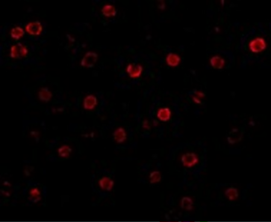
T-cadherin Antibody
| Code | Size | Price |
|---|
| PSI-3583-0.02mg | 0.02mg | £150.00 |
Quantity:
| PSI-3583-0.1mg | 0.1mg | £449.00 |
Quantity:
Prices exclude any Taxes / VAT
Overview
Host Type: Rabbit
Antibody Isotype: IgG
Antibody Clonality: Polyclonal
Regulatory Status: RUO
Applications:
- Enzyme-Linked Immunosorbent Assay (ELISA)
- Immunofluorescence (IF)
- Western Blot (WB)
Images
Documents
Further Information
Additional Names:
T-cadherin Antibody: HB6, CD39L3, NTPDase-3, Ectonucleoside triphosphate diphosphohydrolase 3, CD39 antigen-like 3, NTPDase 3
Application Note:
T-cadherin antibody can be used for the detection of T-cadherin by Western blot at 0.5 - 1 μg/mL. Antibody can also be used for for immunohistochemistry starting at 5 μg/mL and immunocytochemistry starting at 20 μg/mL. For immunofluorescence start at 20 μg/mL.
Antibody validated: Western Blot in mouse samples; Immunohistochemistry in mouse samples and Immunofluorescence in human and mouse samples. All other applications and species not yet tested.
Antibody validated: Western Blot in mouse samples; Immunohistochemistry in mouse samples and Immunofluorescence in human and mouse samples. All other applications and species not yet tested.
Background:
T-cadherin Antibody: T-cadherin was initially identified as cadherin-type cell adhesion molecule expressed in various neuronal populations in a temporally and spatially restricted pattern during axon growth. T-cadherin is an atypical member of the cadherin family because it does not possess the typical transmembrane and cytoplasmic domains but is instead anchored to the plasma membrane by glycosylphosphatidylinositol (GPI) linkage. T-cadherin may play a role in malignant tumor development as loss of the chromosome locus containing the T-cadherin gene correlates with the development of a variety of cancers. Recently it has been shown that T-cadherin can act as a receptor for hexameric and high-molecular weight forms of adiponectin, suggesting that T-cadherin may also play a role in metabolic regulation.
Background References:
- Ranscht B and Dours-Zimmerman MT. T-cadherin, a novel cadherin cell adhesion molecule in the nervous system lacks the conserved cytoplasmic region. Neuron 1991; 7:391-402.
- Ivanov DB, Philippova MP, and Tkachuk VA. Structure and functions of classical cadherins Biochemistry (Moscow) 2001; 66:1175-66.
- Takeuchi T, Misaki A, Chen BK, et al. H-cadherin expression in breast cancer. Histopathology 1999; 35:87-88.
- Sato M, Mori Y, Sakurada A, et al. The H-cadherin (CDH13) gene is inactivated in human lung cancer. Hum. Gen. 1998; 103:96-101.
Buffer:
T-cadherin Antibody is supplied in PBS containing 0.02% sodium azide.
Concentration:
1 mg/mL
Conjugate:
Unconjugated
DISCLAIMER:
Optimal dilutions/concentrations should be determined by the end user. The information provided is a guideline for product use. This product is for research use only.
Homology:
Predicted species reactivity based on immunogen sequence: Bovine: (93%)
Immunogen:
T-cadherin antibody was raised against a 15 amino acid synthetic peptide from near the amino terminus of human T-cadherin.
The immunogen is located within amino acids 150 - 200 of T-cadherin.
The immunogen is located within amino acids 150 - 200 of T-cadherin.
NCBI Gene ID #:
956
NCBI Official Name:
ectonucleoside triphosphate diphosphohydrolase 3
NCBI Official Symbol:
ENTPD3
NCBI Organism:
Homo sapiens
Physical State:
Liquid
PREDICTED MOLECULAR WEIGHT:
Predicted: 78, 84 kDa
Observed: 85 kDa
Observed: 85 kDa
Protein Accession #:
NP_001248
Protein GI Number:
166197701
Purification:
T-cadherin Antibody is affinity chromatography purified via peptide column.
Research Area:
Signal Transduction
SPECIFICITY:
At least two isoforms of T-cadherin are known to exist.
Swissprot #:
O75355
User NOte:
Optimal dilutions for each application to be determined by the researcher.
Related Products
| Product Name | Product Code | Supplier | T-cadherin Peptide | PSI-3583P | ProSci | Summary Details | |||||||||||||||||||||||||||||||||||||||||||||||||||||||||||||||||||||||||||||||||||||||||||||
|---|---|---|---|---|---|---|---|---|---|---|---|---|---|---|---|---|---|---|---|---|---|---|---|---|---|---|---|---|---|---|---|---|---|---|---|---|---|---|---|---|---|---|---|---|---|---|---|---|---|---|---|---|---|---|---|---|---|---|---|---|---|---|---|---|---|---|---|---|---|---|---|---|---|---|---|---|---|---|---|---|---|---|---|---|---|---|---|---|---|---|---|---|---|---|---|---|---|---|---|


