TSLP Antibody
| Code | Size | Price |
|---|
| PSI-4023-0.02mg | 0.02mg | £150.00 |
Quantity:
| PSI-4023-0.1mg | 0.1mg | £449.00 |
Quantity:
Prices exclude any Taxes / VAT
Overview
Host Type: Rabbit
Antibody Isotype: IgG
Antibody Clonality: Polyclonal
Regulatory Status: RUO
Applications:
- Enzyme-Linked Immunosorbent Assay (ELISA)
- Immunofluorescence (IF)
- Immunohistochemistry (IHC)
- Western Blot (WB)
Images
Documents
Further Information
Additional Names:
TSLP Antibody: Thymic stromal lymphopoietin
Application Note:
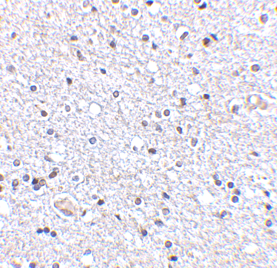
WB: 0.25 - 4 μg/mL; IHC-P: 2.5 μg/mL; IF: 20 μg/mL.
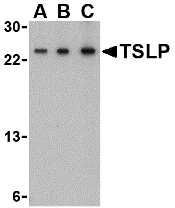
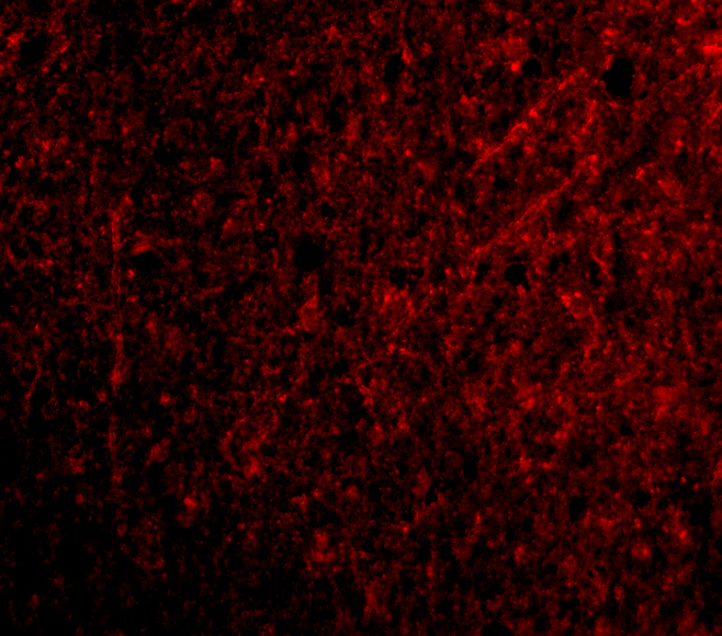
Antibody validated: Western Blot in human and mouse samples; Immunohistochemistry and Immunofluorescence in human and mouse samples; Immunocytochemistry in human samples. All other applications and species not yet tested.
Antibody validated: Western Blot in human and mouse samples; Immunohistochemistry and Immunofluorescence in human and mouse samples; Immunocytochemistry in human samples. All other applications and species not yet tested.
Background:
TSLP Antibody: Thymic stromal lymphopoietin (TSLP) has recently been identified as an important factor capable of driving dendritic cell maturation and activation. TSLP is a four-helix-bundle cytokine that is expressed mainly by barrier epithelial cells and is a potent activator of several cell types such as myeloid dendritic cells. TSLP is involved in the positive selection of regulatory T cells, maintenance of peripheral CD4+ T cell homeostasis and the induction of CD4+ T cell-mediated allergic reaction. TSLP is also capable of supporting the growth of fetal liver and adult B cell progenitors and their differentiation to the IgM-positive stage of B cell development. Amino acid sequence analysis has shown poor homology between human and mouse TSLP although they exhibit similar biological functions and are expressed in similar tissues. At least two differentially spliced isoforms of TSLP are known to exist.
Background References:
- Ziegler and Liu. Nature Immunol. 2006; 7:709-14.
- Sims et al. J. Exp. Med. 2000; 192:671-80.
- Levin et al. J. Immunol. 1999; 162:677-83.
- Quentmeier et al. Leukemia 2001; 15:1286-92.
Buffer:
TSLP Antibody is supplied in PBS containing 0.02% sodium azide.
Concentration:
1 mg/mL
Conjugate:
Unconjugated
DISCLAIMER:
Optimal dilutions/concentrations should be determined by the end user. The information provided is a guideline for product use. This product is for research use only.
Immunogen:
Anti-TSLP antibody (4023) was raised against a peptide corresponding to 19 amino acids near the center of human TSLP.
The immunogen is located within the amino acids 40-90 of TSLP.
The immunogen is located within the amino acids 40-90 of TSLP.
ISOFORMS:
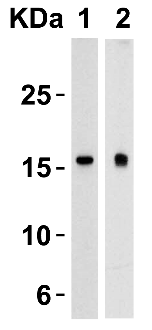
Human TSLP has 2 isoforms, including isoform 1 (159aa, 18kD) and isoform 2 (63aa, 7kD). Mouse TSLP has one isoform (140aa, 16kD) and Rat TSLP also has one isoform (135aa, 16kD). 4023 can detect only longer human isoform, but cannot detect shorter human isoform, mouse and rat.
NCBI Gene ID #:
85480
NCBI Official Name:
thymic stromal lymphopoietin
NCBI Official Symbol:
TSLP
NCBI Organism:
Homo sapiens
Physical State:
Liquid
PREDICTED MOLECULAR WEIGHT:
Predicted: 18 kD
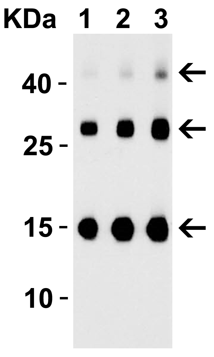
Observed: 17, 24 kD (Post-modifications: 2 N linked glycosylations)
Observed: 17, 24 kD (Post-modifications: 2 N linked glycosylations)
Protein Accession #:
NP_149024
Protein GI Number:
14719428
Purification:
TSLP Antibody is affinity chromatography purified via peptide column.
Research Area:
Immunology
Swissprot #:
Q969D9
User NOte:
Optimal dilutions for each application to be determined by the researcher.
VALIDATION:
Recombinant Protein Test (Figure 2): Anit-TSLP antibodies (4023) detected human TSLP recombinant protein at different concentrations.
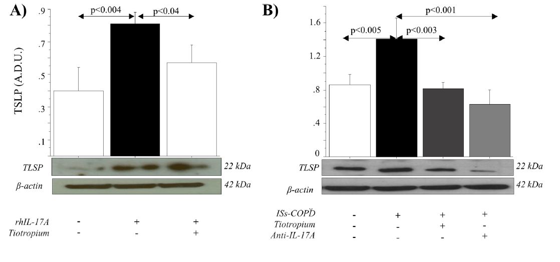
Regulated Expression Validation (Figure 8-11): Anit-TSLP antibodies (4021) detected increased expression in multiple tissue or cells after treatment with cytokines or chemicals, in which the increase in the expression was inhibited by co-treatment with anti-cholinergic drugs (Figure 8).
References
- Anzalone et al. IL-17A-associated IKK-? signaling induced TSLP production in epithelial cells of COPD patients. Exp Mol Med. 2018;50(10):131. PMID: PMID: 30291224.
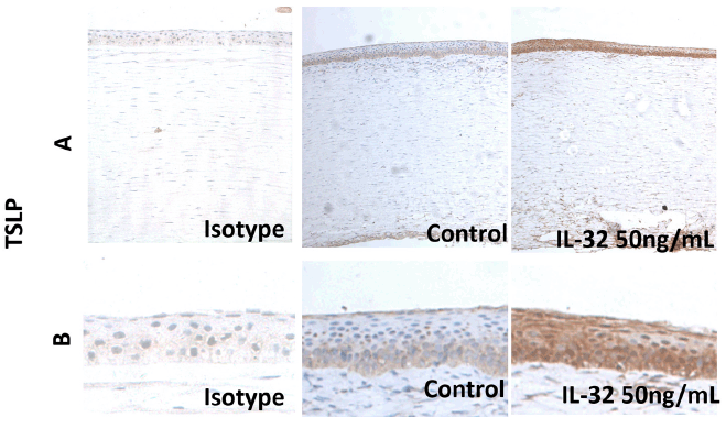
- Lin et al. Interleukin-32 induced thymic stromal lymphopoietin plays a critical role in the inflammatory response in human corneal epithelium. Cell Signal. 2018;49:39-45. PMID: PMID: 29803543.
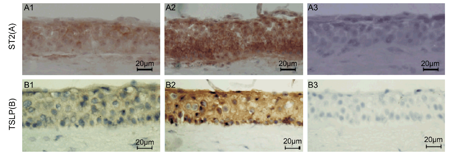
- Lin et al. Regulation of interleukin 33/ST2 signaling of human corneal epithelium in allergic diseases. Int J Ophthalmol. 2013;6(1):23-9. PMID: PMID: 23550226.
- Allakhverdi et al. Mast Cell-Activated Bone Marrow Mesenchymal Stromal Cells Regulate Proliferation and Lineage Commitment of CD34(+) Progenitor Cells. Front Immunol. 2013;4:461. PMID: PMID: 24381572.
- Li et al. Short ragweed pollen triggers allergic inflammation through Toll-like receptor 4-dependent thymic stromal lymphopoietin/OX40 ligand/OX40 signaling pathways. J Allergy Clin Immunol. 2011;128(6):1318-1325.e2. PMID: PMID: 21820713.
- Zheng et al. TSLP and downstream molecules in experimental mouse allergic conjunctivitis. Invest Ophthalmol Vis Sci. 2010;51(6):3076-82. PMID: PMID: 20107175.
- Ma et al. Human corneal epithelium-derived thymic stromal lymphopoietin links the innate and adaptive immune responses via TLRs and Th2 cytokines. Invest Ophthalmol Vis Sci. 2009;50(6):2702-9. PMID: PMID: 19151401.
- Cui et al. TSLP Protects Corneas From Pseudomonas aeruginosa Infection by Regulating Dendritic Cells and IL-23-IL-17 Pathway. Invest Ophthalmol Vis Sci. 2018;59(10):4228-4237. PMID: PMID: 30128494.
- Giulia Anzalone. IL-17A induces chromatin remodeling promoting IL-8 and TSLP release in bronchial epithelial cells. Effect of Tiotropium. University of Palermo. PhD Thesis. 2015. PMID:
- Soma Tripathi. Reduced proliferation and increased TSLP expression by lung fibroblasts from COPD patients. University of Manitoba. MSc Thesis. 2013.PMID:
Related Products
| Product Name | Product Code | Supplier | TSLP Peptide | PSI-4023P | ProSci | Summary Details | |||||||||||||||||||||||||||||||||||||||||||||||||||||||||||||||||||||||||||||||||||||||||||||
|---|---|---|---|---|---|---|---|---|---|---|---|---|---|---|---|---|---|---|---|---|---|---|---|---|---|---|---|---|---|---|---|---|---|---|---|---|---|---|---|---|---|---|---|---|---|---|---|---|---|---|---|---|---|---|---|---|---|---|---|---|---|---|---|---|---|---|---|---|---|---|---|---|---|---|---|---|---|---|---|---|---|---|---|---|---|---|---|---|---|---|---|---|---|---|---|---|---|---|---|


