SCF Antibody

ProSci
Product Code: PSI-5165
Product Group: Primary Antibodies
Supplier: ProSci
CodeSizePrice
PSI-5165-0.02mg0.02mg£150.00
Quantity:
PSI-5165-0.1mg0.1mg£449.00
Quantity:
Prices exclude any Taxes / VAT

Overview

Host Type: Rabbit
Antibody Isotype: IgG
Antibody Clonality: Polyclonal
Regulatory Status: RUO
Applications:
  • Enzyme-Linked Immunosorbent Assay (ELISA)
  • Immunofluorescence (IF)
  • Immunohistochemistry (IHC)
  • Western Blot (WB)

Images

1 / 8
<strong>Figure 1 Western Blot Validation in Cell Lines and Tissues of Human, Mouse and Rat</strong><br>
Loading: 15 μg of lysates per lane.
Antibodies: SCF 5165 (1 μg/mL),  1h incubation at RT  in 5% NFDM/TBST.
Secondary: Goat anti-rabbit IgG HRP conjugate at 1:10000 dilution.
2 / 8
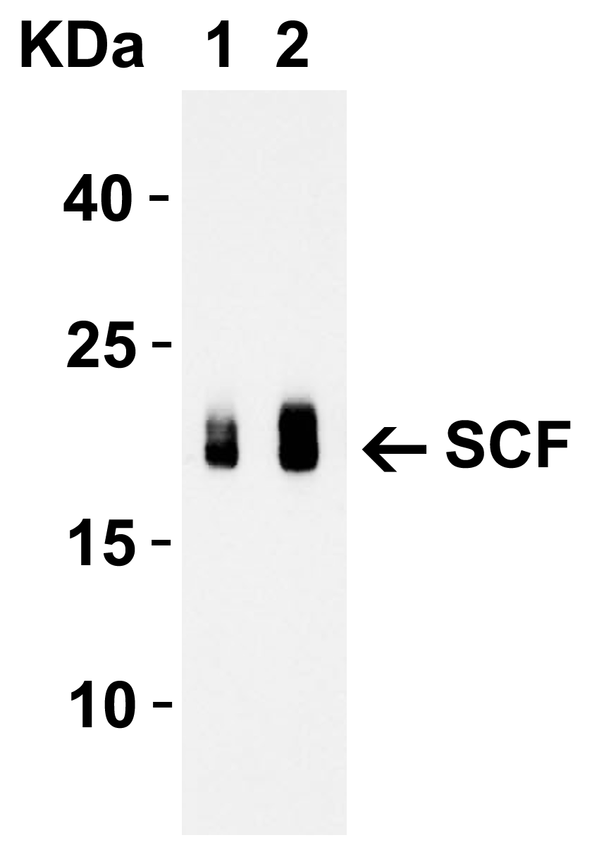
<strong>Figure 2 Western Blot Validation with Recombinant Protein</strong><br>
Loading: 30 ng of human SCF recombinant protein per lane.
Antibodies: SCF 5165 (Lane 1: 1 μg/mL and Lane 2: 2 μg/mL), 1h incubation at RT  in 5% NFDM/TBST.
Secondary: Goat anti-rabbit IgG HRP conjugate at 1:10000 dilution.
Observed at around 20kD.
3 / 8
<strong>Figure 3 Western Blot Validation in Human HeLa Cell Lines</strong><br>
Loading: 15 μg of lysates per lane.
Antibodies: SCF 5165 (2 μg/mL), 1h incubation at RT in 5% NFDM/TBST.
Secondary: Goat anti-rabbit IgG HRP conjugate at 1:10000 dilution.
4 / 8
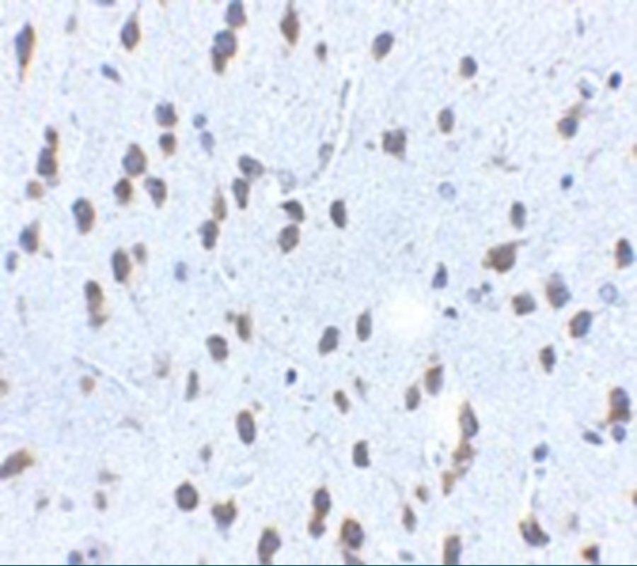
<strong>Figure 4  Immunohistochemistry Validation of SCF in Mouse Brain Tissue </strong><br> 
Immunohistochemical analysis of paraffin-embedded mouse brain tissue using anti-SCF antibody (5165) at 2.5 μg/ml. Tissue was fixed with formaldehyde and blocked with 10% serum for 1 h at RT; antigen retrieval was by heat mediation with a citrate buffer (pH6). Samples were incubated with primary antibody overnight at 4 ˚C. A goat anti-rabbit IgG H&L (HRP) at 1/250 was used as secondary. Counter stained with Hematoxylin.
5 / 8
<strong>Figure  5 Immunofluorescence Validation of SCF in Human Brain Tissue</strong><br>
Immunofluorescent analysis of 4% paraformaldehyde-fixed human brain tissue labeling SCF with 5165 at 20 μg/mL, followed by goat anti-rabbit IgG secondary antibody at 1/500 dilution (red).
6 / 8
<strong>Figure 6  Regulation Validation of SCF in Streptozotocin (STZ)-induced Diabetic Mice (Zhang et al.,  2016) </strong><br>
WB analysis showed protein expression level of SCF detected by anti-SCF antibody (5165) in gastric smooth muscle was  significantly decreased in STZ-induced diabetic mice as compared to the control.
7 / 8
<strong>Figure 7  Regulated Expression Validation of SCF in Gastric Smooth Muscle Cells (GSMCs) of Mice  (Zhang et al., 2016) </strong><br>
WB analysis of protein expression level of SCF detected by anti-SCF antibody (5165). GSMCs were treated with PI3K inhibitor (LY294002), AT1R inhibitor (ZD7155) and AT2R inhibitor (PD123319) before Ang II treatment. Ang II (10<sup>-8</sup>mol/L) significantly increased SCF protein expression, which was reduced by treatment of LY294002 and ZD7155.
8 / 8
<strong>Figure 8  Regulated Expression Validation of SCF in GSMCs of Normal Mice  (Wu et al., 2013) </strong><br>
WB analysis of protein expression level of SCF detected by anti-SCF antibody (5165). GSMCs were treated with C-type natriuretic peptide (CNP) at different doses for 48hr. CNP(10<sup>-7</sup>mol/L and 10<sup>-6</sup>mol/L) significantly decreased SCF protein expression level as compared to the control group.

<strong>Figure 1 Western Blot Validation in Cell Lines and Tissues of Human, Mouse and Rat</strong><br>
Loading: 15 μg of lysates per lane.
Antibodies: SCF 5165 (1 μg/mL),  1h incubation at RT  in 5% NFDM/TBST.
Secondary: Goat anti-rabbit IgG HRP conjugate at 1:10000 dilution.
<strong>Figure 2 Western Blot Validation with Recombinant Protein</strong><br>
Loading: 30 ng of human SCF recombinant protein per lane.
Antibodies: SCF 5165 (Lane 1: 1 μg/mL and Lane 2: 2 μg/mL), 1h incubation at RT  in 5% NFDM/TBST.
Secondary: Goat anti-rabbit IgG HRP conjugate at 1:10000 dilution.
Observed at around 20kD.
<strong>Figure 3 Western Blot Validation in Human HeLa Cell Lines</strong><br>
Loading: 15 μg of lysates per lane.
Antibodies: SCF 5165 (2 μg/mL), 1h incubation at RT in 5% NFDM/TBST.
Secondary: Goat anti-rabbit IgG HRP conjugate at 1:10000 dilution.
<strong>Figure 4  Immunohistochemistry Validation of SCF in Mouse Brain Tissue </strong><br> 
Immunohistochemical analysis of paraffin-embedded mouse brain tissue using anti-SCF antibody (5165) at 2.5 μg/ml. Tissue was fixed with formaldehyde and blocked with 10% serum for 1 h at RT; antigen retrieval was by heat mediation with a citrate buffer (pH6). Samples were incubated with primary antibody overnight at 4 ˚C. A goat anti-rabbit IgG H&L (HRP) at 1/250 was used as secondary. Counter stained with Hematoxylin.
<strong>Figure  5 Immunofluorescence Validation of SCF in Human Brain Tissue</strong><br>
Immunofluorescent analysis of 4% paraformaldehyde-fixed human brain tissue labeling SCF with 5165 at 20 μg/mL, followed by goat anti-rabbit IgG secondary antibody at 1/500 dilution (red).
<strong>Figure 6  Regulation Validation of SCF in Streptozotocin (STZ)-induced Diabetic Mice (Zhang et al.,  2016) </strong><br>
WB analysis showed protein expression level of SCF detected by anti-SCF antibody (5165) in gastric smooth muscle was  significantly decreased in STZ-induced diabetic mice as compared to the control.
<strong>Figure 7  Regulated Expression Validation of SCF in Gastric Smooth Muscle Cells (GSMCs) of Mice  (Zhang et al., 2016) </strong><br>
WB analysis of protein expression level of SCF detected by anti-SCF antibody (5165). GSMCs were treated with PI3K inhibitor (LY294002), AT1R inhibitor (ZD7155) and AT2R inhibitor (PD123319) before Ang II treatment. Ang II (10<sup>-8</sup>mol/L) significantly increased SCF protein expression, which was reduced by treatment of LY294002 and ZD7155.
<strong>Figure 8  Regulated Expression Validation of SCF in GSMCs of Normal Mice  (Wu et al., 2013) </strong><br>
WB analysis of protein expression level of SCF detected by anti-SCF antibody (5165). GSMCs were treated with C-type natriuretic peptide (CNP) at different doses for 48hr. CNP(10<sup>-7</sup>mol/L and 10<sup>-6</sup>mol/L) significantly decreased SCF protein expression level as compared to the control group.

Further Information

Additional Names:
SCF Antibody: SF, MGF, SCF, FPH2, KL-1, Kitl, SHEP7, Kit ligand, Mast cell growth factor
Application Note:
WB: 1-2 μg/mL; IHC: 2.5 μg/mL; IF: 20 μg/mL.

Antibody validated: Western Blot in human, mouse and rat samples; Immunohistochemistry in mouse samples; Immunofluorescence in human samples. All other applications and species not yet tested.
Background:
SCF Antibody: Stem cell factor (SCF) is the ligand of the c-Kit oncogene and is expressed by various structural and inflammatory cells in the airways. Binding of SCF by the c-Kit receptor leads to homodimerization of the receptor and the activation of signalling pathways such as PI-3, PLC-gamma, Jak/STAT, and MAP kinase pathways. SCF expression leads to the induction of mast cell survival and the expression and release of histamine, pro-inflammatory cytokines and chemokines. The inhibition of the SCF/c-Kit pathway leads to a decrease in histamine levels, mast cell and eosinophil infiltration, IL-4 production and airway hyperresponsiveness, suggesting this pathway may be a useful therapeutic target in inflammatory diseases such as asthma. At least two isoforms of SCF are known to exist.
Background References:
  • Reber et al. Euro. J. Pharmacology 2006; 533:327-40.
  • Jensen et al. Inflamm. Allergy Drug Targets 2007; 6:57-62.
  • Lukacs et al. J. Immunol. 1996; 156:3945-51.
Buffer:
SCF Antibody is supplied in PBS containing 0.02% sodium azide.
Concentration:
1 mg/mL
Conjugate:
Unconjugated
DISCLAIMER:
Optimal dilutions/concentrations should be determined by the end user. The information provided is a guideline for product use. This product is for research use only.
Homology:
Predicted species reactivity based on immunogen sequence: Bovine: (71%)
Immunogen:
Anti-SCF antibody (5165) was raised against a peptide corresponding to 18 amino acids near the center of human SCF.

The immunogen is located within amino acids 100-150 of SCF.
ISOFORMS:
Human SCF has 3 isoforms, including isoform 1 (273aa, 30.9kD), isoform 2 (245aa, 27.9kD) and isoform 3 (238aa, 26.7kD). Mouse SCF has 2 isoforms, including isoform 1 (273aa, 30.6kD) and isoform 2 (245aa, 27.6kD). Rat SCF also has 2 isoforms, including isoform 1 (273aa, 30.7kD) and isoform 2 (245aa, 27.7kD). 5165 can detect human and possibly detect mouse and rat.
NCBI Gene ID #:
4254
NCBI Official Name:
KIT ligand
NCBI Official Symbol:
KITLG
NCBI Organism:
Homo sapiens
Physical State:
Liquid
PREDICTED MOLECULAR WEIGHT:
Predicted: 31kD

Observed: 36 kD (31kD + 4 N-linked Glycosylations)
Protein Accession #:
P21583
Protein GI Number:
134289
Purification:
SCF Antibody is affinity chromatography purified via peptide column.
Research Area:
Stem Cell
Swissprot #:
P21583
User NOte:
Optimal dilutions for each application to be determined by the researcher.
VALIDATION:

Recombinant Protein Test (Figure 2): Anti-SCF antibodies (5165) detected human SCF recombinant protein at different concentrations.

Regulated expression validation (Figure 7 & 8): SCF expression detected by anti-SCF antibodies (5165) was up-regulated by angiotensin II (Ang II) treatment and was down-regulated by two inhibitors (Figure 7) or CNP treatment (Figure 8).

References

  1. Zhang et al. Up-regulation of the Ang II/AT1 receptor may compensate for the loss of gastric antrum ICC via the PI3k/Akt signaling pathway in STZ-induced diabetic mice. Mol Cell Endocrinol. 2016;423:77-86.PMID: 26773730
  2. Wu et al. Diabetes-induced loss of gastric ICC accompanied by up-regulation of natriuretic peptide signaling pathways in STZ-induced diabetic mice. Peptides. 2013;40:104-11. PMID: 23352981

Related Products

Product NameProduct CodeSupplier 
SCF PeptidePSI-5165PProSci Summary Details