RSPO1 Antibody

ProSci
Product Code: PSI-5171
Product Group: Primary Antibodies
Supplier: ProSci
CodeSizePrice
PSI-5171-0.02mg0.02mg£150.00
Quantity:
PSI-5171-0.1mg0.1mg£449.00
Quantity:
Prices exclude any Taxes / VAT

Overview

Host Type: Rabbit
Antibody Isotype: IgG
Antibody Clonality: Polyclonal
Regulatory Status: RUO
Applications:
  • Enzyme-Linked Immunosorbent Assay (ELISA)
  • Immunofluorescence (IF)
  • Immunohistochemistry (IHC)
  • Western Blot (WB)

Images

1 / 8
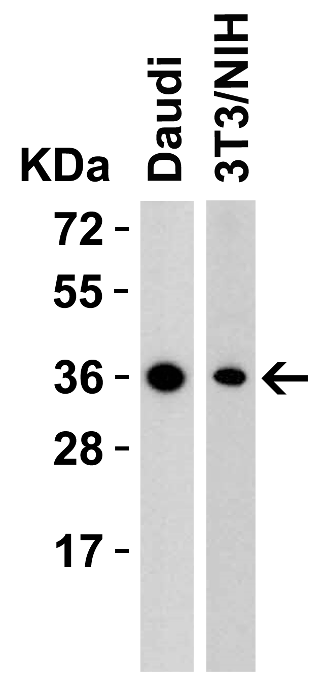
<strong>Figure 1 Western Blot Validation in Human and Mouse Cell Lines</strong><br>
Loading: 15 μg of lysates per lane.
Antibodies: RSPO1, 5171 (2 μg/mL), 1h incubation at RT in 5% NFDM/TBST.
Secondary: Goat anti-rabbit IgG HRP conjugate at 1:10000 dilution.
2 / 8
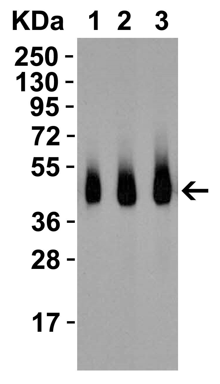
<strong>Figure 2 Western Blot Validation with Recombinant Protein</strong><br>
Loading: 30 ng of human RSPO1 recombinant protein per lane.
Antibodies: RSPO1, 5171 (Lane 1: 0.5 μg/mL, Lane 2: 1 μg/mL,  Lane 3: 2 μg/mL), 1h incubation at RT  in 5% NFDM/TBST.
Secondary: Goat anti-rabbit IgG HRP conjugate at 1:10000 dilution.
3 / 8
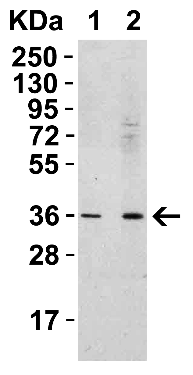
<strong>Figure 3 Western Blot Validation in Human Spleen </strong><br>
Loading: 15 μg of lysates per lane.
Antibodies: RSPO1, 5171 (Lane 1: 0.5 μg/mL, Lane 2: 1 μg/mL), 1h incubation at RT in 5% NFDM/TBST.
Secondary: Goat anti-rabbit IgG HRP conjugate at 1:10000 dilution.
4 / 8
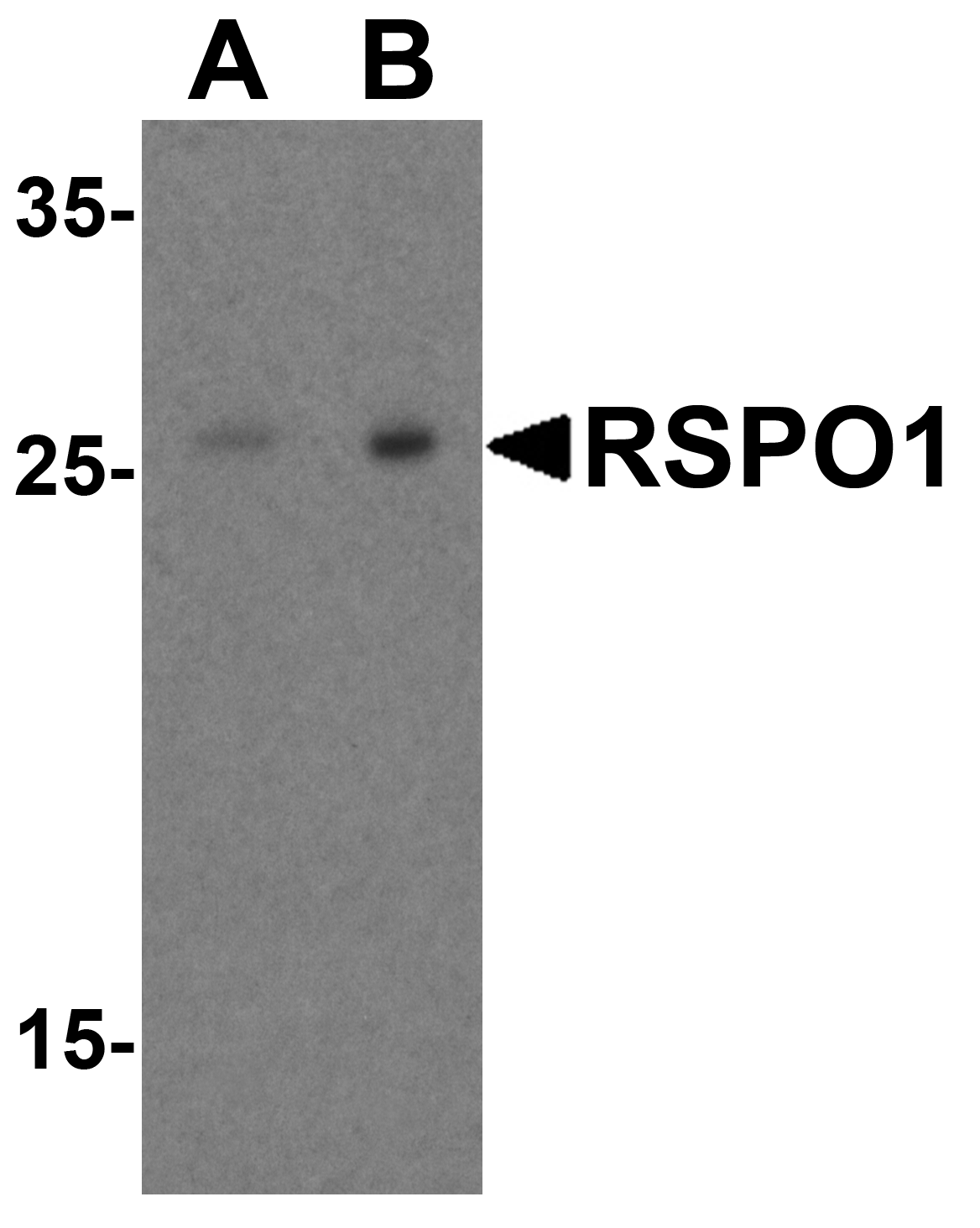
<strong>Figure 4 Western Blot Validation in Human Heart</strong><br>
Loading: 15 μg of lysates per lane.
Antibodies: RSPO1, 5171 (A: 1 μg/mL, B: 2 μg/mL), 1h incubation at RT in 5% NFDM/TBST.
Secondary: Goat anti-rabbit IgG HRP conjugate at 1:10000 dilution.
5 / 8
<strong>Figure 5 Immunofluorescence Validation of RSPO1 in Human Spleen </strong><br>
Immunofluorescent analysis of 4% paraformaldehyde-fixed human spleen labeling RSPO1 with 5171 at 5 μg/mL, followed by goat anti-rabbit IgG secondary antibody at 1/500 dilution (green) with Phylloidin staining (red) and DAPI staining (blue).
6 / 8
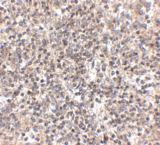
<strong>Figure 6 Immunohistochemistry Validation of RSPO1 in Human Heart </strong><br> 
Immunohistochemical analysis of paraffin-embedded mouse spleen tissue using anti-RSPO1 antibody (5171) at 2.5 μg/ml. Tissue was fixed with formaldehyde and blocked with 10% serum for 1 h at RT; antigen retrieval was by heat mediation with a citrate buffer (pH6). Samples were incubated with primary antibody overnight at 4˚C. A goat anti-rabbit IgG H&L (HRP) at 1/250 was used as secondary. Counter stained with Hematoxylin.
7 / 8
<strong>Figure 7 Immunohistochemistry Validation of RSPO1 in Human Spleen Tissue </strong><br> 
Immunohistochemical analysis of paraffin-embedded human spleen tissue using anti-RSPO1 antibody (5171) at 5 ug/ml. Tissue was fixed with formaldehyde and blocked with 10% serum for 1 h at RT; antigen retrieval was by heat mediation with a citrate buffer (pH6). Samples were incubated with primary antibody overnight at 4˚C. A goat anti-rabbit IgG H&L (HRP) at 1/250 was used as secondary. Counter stained with Hematoxylin.
8 / 8
<strong>Figure 8 Immunohistochemistry Validation of RSPO1 in Human Spleen Tissue </strong><br> 
Immunohistochemical analysis of paraffin-embedded human spleen tissue using anti-RSPO1 antibody (5171) at 2.5 ug/ml. Tissue was fixed with formaldehyde and blocked with 10% serum for 1 h at RT; antigen retrieval was by heat mediation with a citrate buffer (pH6). Samples were incubated with primary antibody overnight at 4˚C. A goat anti-rabbit IgG H&L (HRP) at 1/250 was used as secondary. Counter stained with Hematoxylin.

<strong>Figure 1 Western Blot Validation in Human and Mouse Cell Lines</strong><br>
Loading: 15 μg of lysates per lane.
Antibodies: RSPO1, 5171 (2 μg/mL), 1h incubation at RT in 5% NFDM/TBST.
Secondary: Goat anti-rabbit IgG HRP conjugate at 1:10000 dilution.
<strong>Figure 2 Western Blot Validation with Recombinant Protein</strong><br>
Loading: 30 ng of human RSPO1 recombinant protein per lane.
Antibodies: RSPO1, 5171 (Lane 1: 0.5 μg/mL, Lane 2: 1 μg/mL,  Lane 3: 2 μg/mL), 1h incubation at RT  in 5% NFDM/TBST.
Secondary: Goat anti-rabbit IgG HRP conjugate at 1:10000 dilution.
<strong>Figure 3 Western Blot Validation in Human Spleen </strong><br>
Loading: 15 μg of lysates per lane.
Antibodies: RSPO1, 5171 (Lane 1: 0.5 μg/mL, Lane 2: 1 μg/mL), 1h incubation at RT in 5% NFDM/TBST.
Secondary: Goat anti-rabbit IgG HRP conjugate at 1:10000 dilution.
<strong>Figure 4 Western Blot Validation in Human Heart</strong><br>
Loading: 15 μg of lysates per lane.
Antibodies: RSPO1, 5171 (A: 1 μg/mL, B: 2 μg/mL), 1h incubation at RT in 5% NFDM/TBST.
Secondary: Goat anti-rabbit IgG HRP conjugate at 1:10000 dilution.
<strong>Figure 5 Immunofluorescence Validation of RSPO1 in Human Spleen </strong><br>
Immunofluorescent analysis of 4% paraformaldehyde-fixed human spleen labeling RSPO1 with 5171 at 5 μg/mL, followed by goat anti-rabbit IgG secondary antibody at 1/500 dilution (green) with Phylloidin staining (red) and DAPI staining (blue).
<strong>Figure 6 Immunohistochemistry Validation of RSPO1 in Human Heart </strong><br> 
Immunohistochemical analysis of paraffin-embedded mouse spleen tissue using anti-RSPO1 antibody (5171) at 2.5 μg/ml. Tissue was fixed with formaldehyde and blocked with 10% serum for 1 h at RT; antigen retrieval was by heat mediation with a citrate buffer (pH6). Samples were incubated with primary antibody overnight at 4˚C. A goat anti-rabbit IgG H&L (HRP) at 1/250 was used as secondary. Counter stained with Hematoxylin.
<strong>Figure 7 Immunohistochemistry Validation of RSPO1 in Human Spleen Tissue </strong><br> 
Immunohistochemical analysis of paraffin-embedded human spleen tissue using anti-RSPO1 antibody (5171) at 5 ug/ml. Tissue was fixed with formaldehyde and blocked with 10% serum for 1 h at RT; antigen retrieval was by heat mediation with a citrate buffer (pH6). Samples were incubated with primary antibody overnight at 4˚C. A goat anti-rabbit IgG H&L (HRP) at 1/250 was used as secondary. Counter stained with Hematoxylin.
<strong>Figure 8 Immunohistochemistry Validation of RSPO1 in Human Spleen Tissue </strong><br> 
Immunohistochemical analysis of paraffin-embedded human spleen tissue using anti-RSPO1 antibody (5171) at 2.5 ug/ml. Tissue was fixed with formaldehyde and blocked with 10% serum for 1 h at RT; antigen retrieval was by heat mediation with a citrate buffer (pH6). Samples were incubated with primary antibody overnight at 4˚C. A goat anti-rabbit IgG H&L (HRP) at 1/250 was used as secondary. Counter stained with Hematoxylin.

Further Information

Additional Names:
RSPO1 Antibody: RSPO, CRISTIN3, R-spondin-1, Roof plate-specific spondin-1, hRspo1
Application Note:
WB: 0.5-2 μg/mL; IHC: 2.5-5 μg/mL; IF: 5 μg/mL.

Antibody validated: Western Blot in human and mouse samples; Immunohistochemistry in human samples; Immunofluorescence in human samples. All other applications and species not yet tested.
Background:
RSPO1 Antibody: RSPO1 is a member of a family of secreted growth factors that can operate through the canonical Wnt signaling pathway by stabilizing the intracellular beta-catenin, thereby regulating functions mediated by beta-catenin such as cell fate decisions and embryonic patterning. RSPO1 was recently identified through linkage analysis to be involved in sex determination and mammalian ovarian development. RSPO1 is thought to regulate cellular responsiveness to Wnt ligands by modulating the cell-surface expression of the Wnt co-receptor LRP6 by interfering with the DKK/Kremen-mediated internalization of LRP6 through an interaction with Kremen, resulting in increased LRP6 cell-surface levels.
Background References:
  • Kamata et al. Biochim. Biophys. Acta 2004; 1676:51-61.
  • Kim et al. Cell Cycle 2006; 5:23-6.
  • Parma et al. Nat. Genet. 2006; 38:1304-9.
  • Chassot et al. Hum. Mol. Genet. 2008; 17:1278-91.
Buffer:
RSPO1 Antibody is supplied in PBS containing 0.02% sodium azide.
Concentration:
1 mg/mL
Conjugate:
Unconjugated
DISCLAIMER:
Optimal dilutions/concentrations should be determined by the end user. The information provided is a guideline for product use. This product is for research use only.
Homology:
Predicted species reactivity based on immunogen sequence: Rat (100%).
Immunogen:
Anti-RSPO1 antibody (5171) was raised against a peptide corresponding to 16 amino acids near the amino terminus of human RSPO1.

The immunogen is located within the first 50 amino acids of RSPO1.
ISOFORMS:
Human RSPO1 has 3 isoforms, including isoform 1 (263aa, 29kD), isoform 2 (236aa, 26kD), and isoform 3 (200aa, 22kD). Mouse RSPO1 had 1 isoform (265aa, 29kD) and rat RSPO1 had 1 isoform (262aa, 29kD). 5171 can detect human, mouse and rat.
NCBI Gene ID #:
284654
NCBI Official Name:
R-spondin homolog (Xenopus laevis)
NCBI Official Symbol:
RSPO1
NCBI Organism:
Homo sapiens
Physical State:
Liquid
PREDICTED MOLECULAR WEIGHT:
Predicted: 29kD

Observed: 36 kD (Post-modification: 1 N-linked glycosylation)
Protein Accession #:
NP_001033722
Protein GI Number:
84490388
Purification:
RSPO1 Antibody is affinity chromatography purified via peptide column.
Research Area:
Signal Transduction
SPECIFICITY:
At least three isoforms of RSPO1 are known to exist; this antibody will detect the largest and smallest isoforms. RSPO1 antibody will not cross-react with other RSPO family members.
Swissprot #:
Q2MKA7
User NOte:
Optimal dilutions for each application to be determined by the researcher.
VALIDATION:

Recombinant Protein Test (Figure 2): Anti-RSPO1 antibodies (5171) detected human RSPO1 recombinant protein at different concentrations.

Related Products

Product NameProduct CodeSupplier 
RSPO1 PeptidePSI-5171PProSci Summary Details