
PD-L1 Antibody

| Code | Size | Price |
|---|
| PSI-46-156-0.1mg | 0.1mg | £626.00 |
Quantity:
Prices exclude any Taxes / VAT
Overview
Host Type: Caprine (Goat)
Antibody Clonality: Polyclonal
Regulatory Status: RUO
Target Species: Human
Applications:
- Enzyme-Linked Immunosorbent Assay (ELISA)
- Immunohistochemistry (IHC)
- Western Blot (WB)
Storage:
Aliquot and store at -20˚C. Minimize freezing and thawing.
 staining of Human Heart (A) lysate + Blocking peptide (B) (35ug protein in RIPA buffer). Detected by chemiluminescence.")
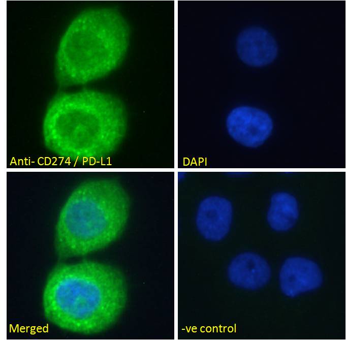
 followed by Alexa Fluor 488 secondary antibody (2ug/ml), showing cytoplasmic staining. The nuclear stain is DAPI (b")
 followed by Alexa Fluor 488 secondary antibody (2ug/ml), showing cytoplasmic staining. The nuclear stain is DAPI (b")
, permeabilized with 0.5% Triton. Primary incubation 1hr (10ug/ml) followed by Alexa Fluor 488 secondary antibody (1ug/ml). IgG control: Unimmunized goat IgG (black line) f")

Documents
Further Information
Additional Names:
CD274, CD274 antigen, PD-L1, PDCD1LG1, B7-H, B7H1, PDL1, PDCD1L1, programmed cell death 1 ligand 1, PDL1, HGNC:17635, CD274 molecule, MGC142294, MGC142296
Application Note:
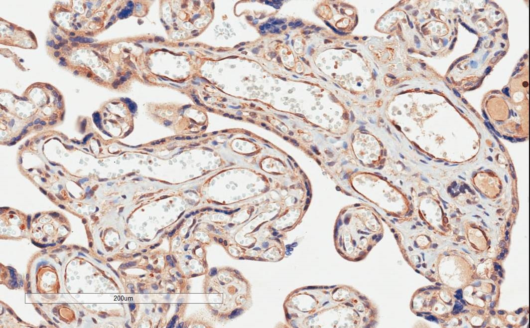
Peptide ELISA: antibody detection limit dilution 1:128000.Western Blot:Approx 40kDa band observed in Human Heart lysates. The observed molecular weight corresponds to glycosylation (calculated MW of 33.3kDa according to NP_054862.1). An additional band was also consistently observed at 55kDa, and was successfully blocked by incubation with the immunizing peptide. Preliminary testing also showed the 37+55kDa bands in lysates of cell lines, A549, Daudi, HeLa, HepG2 and Jurkat Recommended concentration: 0.03-0.1ug/ml. Primary incubation 1 hour at room temperature.Immunohistochemistry:In paraffin embedded Human Placenta shows membranous staining of cytotrophoblasts. Recommended concentration: 2-4ug/ml.Immunofluorescence: Strong expression of the protein seen in the cytoplasm of A431 and U2OS cells. Recommended concentration: 10ug/ml.
Flow Cytometry: Flow cytometric analysis of Jurkat cells. Recommended concentration: 10ug/ml.
Background References:
- Thompson RH, Gillett MD, Cheville JC, Lohse CM, Dong H, Webster WS, Krejci KG, Lobo JR, Sengupta S, Chen L, Zincke H, Blute ML, Strome SE, Leibovich BC, Kwon ED. Costimulatory B7-H1 in renal cell carcinoma patients: Indicator of tumor aggressiveness and potential therapeutic target. Proc Natl Acad Sci USA. 2004 Dec 7;101(49):17174-9. Epub 2004 Dec 7.
Buffer:
Supplied at 0.5 mg/ml in Tris saline, 0.02% sodium azide, pH7.3 with 0.5% bovine serum albumin.
Aliquot and store at -20?C. Minimize freezing and thawing.
Concentration:
500 ug/mL
Conjugate:
Unconjugated
DISCLAIMER:
Optimal dilutions/concentrations should be determined by the end user. The information provided is a guideline for product use. Optimal dilutions/concentrations should be determined by the end user. The information provided is a guideline for product use. This product is for research use only.
Immunogen:
The immunogen for this antibody is: CKKQSDTHLEET
NCBI Gene ID #:
29126
NCBI Official Name:
CD274 molecule
NCBI Official Symbol:
CD274
NCBI Organism:
Homo sapiens
Physical State:
Liquid
Protein Accession #:
NP_054862.1, NP_001254635.1
Protein GI Number:
7661534
Purification:
Purified from goat serum by ammonium sulphate precipitation followed by antigen affinity chromatography using the immunizing peptide.
Research Area:
Cancer
SPECIFICITY:
This antibody is expected to recognize reported isoforms a and b (NP_054862.1; NP_001254635.1) only.
Swissprot #:
Q9NZQ7