SAMHD1 Antibody

ProSci
Product Code: PSI-8007
Product Group: Primary Antibodies
Supplier: ProSci
CodeSizePrice
PSI-8007-0.02mg0.02mg£150.00
Quantity:
PSI-8007-0.1mg0.1mg£449.00
Quantity:
Prices exclude any Taxes / VAT

Overview

Host Type: Rabbit
Antibody Isotype: IgG
Antibody Clonality: Polyclonal
Antibody Clone: 38016914
Regulatory Status: RUO
Target Species:
  • Human
  • Mouse
Applications:
  • Enzyme-Linked Immunosorbent Assay (ELISA)
  • Western Blot (WB)
Storage:
SAMHD1 antibody can be stored at 4˚C for three months and -20˚C, stable for up to one year.

Images

1 / 9
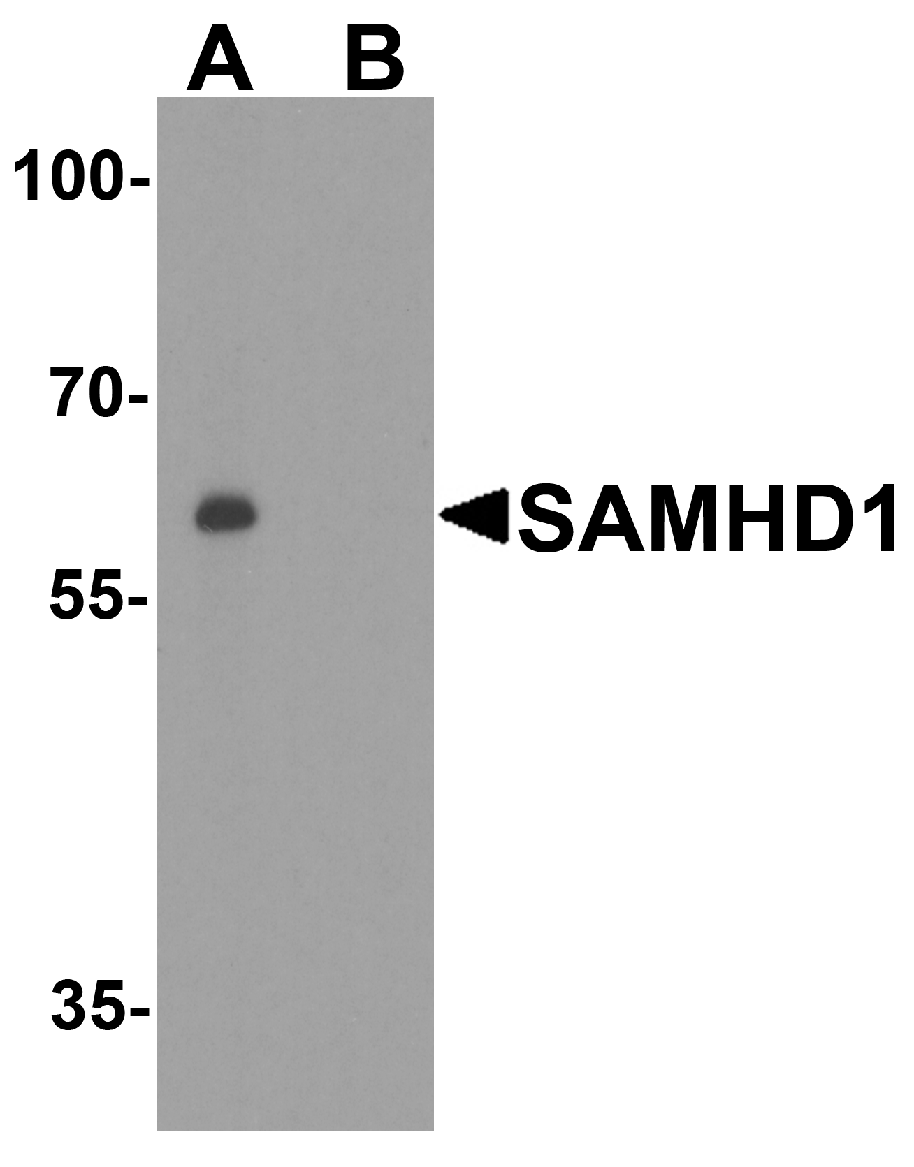
<strong>Figure 1 Western Blot Validation in Human Daudi Cell Lines</strong><br>
Loading: 15 μg of lysates per lane.
Antibodies: SAMHD1 8007, 1 μg/mL, in (A: the absence and B: the presence of blocking peptide), (1h incubation at RT in 5% NFDM/TBST.
Secondary: Goat anti-rabbit IgG HRP conjugate at 1:10000 dilution.
2 / 9
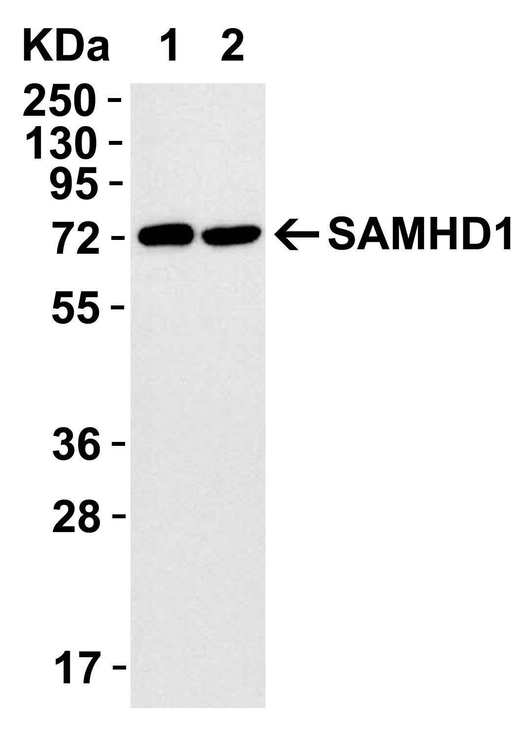
<strong>Figure 2 Overexpression Validation in 293T Transfected Cells</strong><br>
Loading: 15 μg of lysates per lane.
Antibodies: SAMHD1 8007 (0.1 μg/mL), (1h incubation at RT in 5% NFDM/TBST.
Secondary: Goat anti-rabbit IgG HRP conjugate at 1:10000 dilution.
293 cells were transfected with (1) wild-type SAMHD1 or (2) SAMHD1 (mutation T592A).
3 / 9
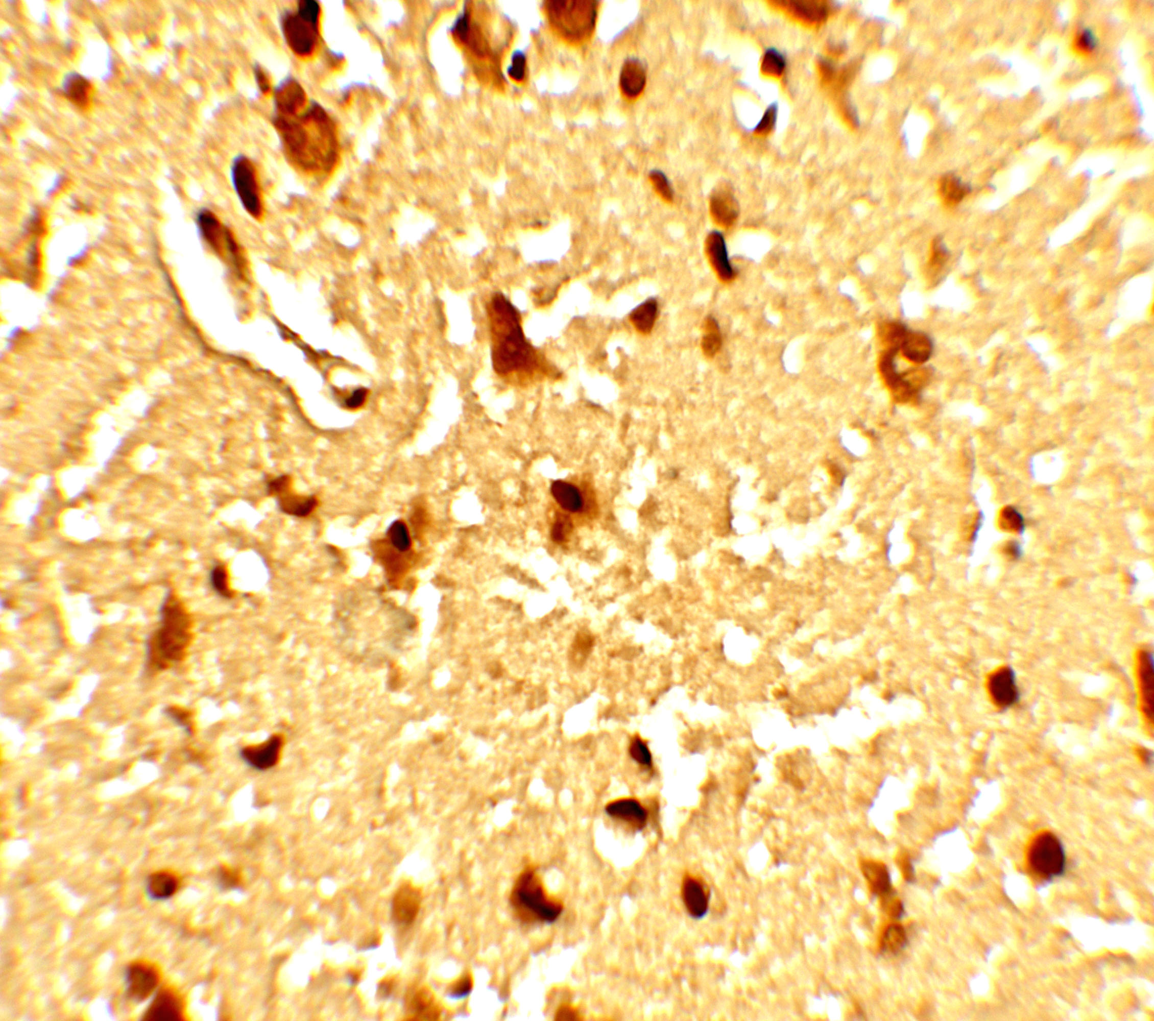
<strong>Figure 3 Immunohistochemistry Validation of SAMHD1 in Human Brain Tissue </strong><br> 
Immunohistochemical analysis of paraffin-embedded Human Brain Tissue using anti- SAMHD1 antibody (8007) at 5 μg/ml. Tissue was fixed with formaldehyde and blocked with 10% serum for 1 h at RT; antigen retrieval was by heat mediation with a citrate buffer (pH6). Samples were incubated with primary antibody overnight at 4 ˚C. A goat anti-rabbit IgG H&L (HRP) at 1/250 was used as secondary. Counter stained with Hematoxylin.
4 / 9
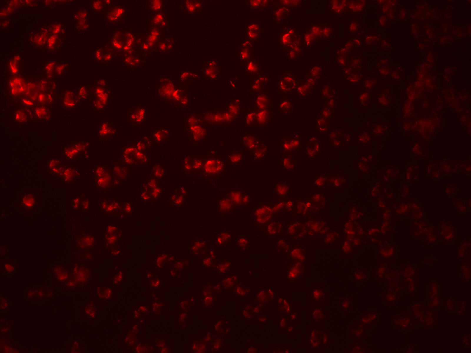
<strong>Figure 4 Immunofluorescence Validation of SAMHD1 in Human Daudi cells</strong><br>
Immunofluorescent analysis of 4% paraformaldehyde-fixed Human Daudi Cells labeling SAMHD1 with 8007 at 20 μg/mL, followed by goat anti-rabbit IgG secondary antibody at 1/500 dilution (red).
5 / 9
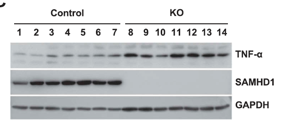
<strong>Figure 5  KO Validation of SAMHD1 in xenograft mice (Kodigepalli et al., 2018)</strong><br> 
 THP-1 control and SAMHD1 KO (THP-1/KO) cells were injected into NSG (non-obese diabetic/severe combined
immune deficient-gamma) mice.  Protein expression levels of SAMHD1 were examined by Western blot with anti-SAMHD1 antibodies (8007) and SAMHD1 was not detected in THP-1/KO cells.
6 / 9
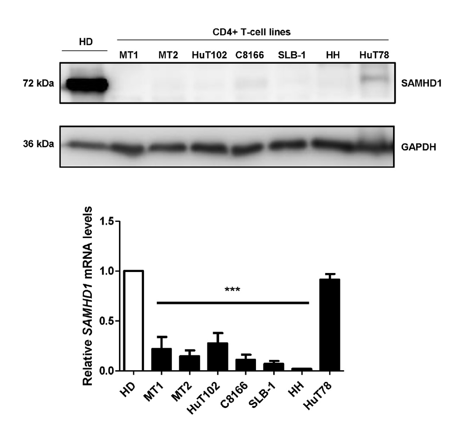
<strong>Figure 6 Overexpression Validation of SAMHD1 in CD4+ T-cells from a healthy donor and transformed CD4+ T-cell lines (Kohnken et al.,  2017) </strong><br>
MT1, MT2, SLB-1, and C8166 were from leukemia patients and HH, HuT78, and HuT102  were from cutaneous T-cell lymphoma (CTCL) patients.  SAMHD1 protein expression detected by anti-SAMHD1 antibodies (8007) was significantly increased in normal CD4+ T-cells as compared to leukemia- and CTCL- derived CD4+ T-cell lines. This blot is representative from four independent experiments with four healthy donors.
7 / 9
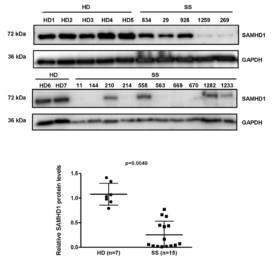
<strong>Figure 7 Overexpression  of SAMHD1 in CD4+ T-cells from Healthy Donors and S?zary Syndrome (SS) patients
(Kohnken et al.,  2017)</strong><br>
SAMHD1 protein expression detected by anti-SAMHD1 antibodies (8007) was significantly reduced in CD+ T-cells from 15 SS patients as compared to those from 7 healthy donors.
8 / 9
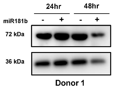
<strong>Figure 8 Regulated Expression Validation of SAMHD1 in CD4+ T-cells from a healthy donor (Kohnken et al.,  2017)</strong><br>
SAMHD1 protein expression detected by anti-SAMHD1 antibodies (8007) was significantly decreased by about 40% relative to control cells at 48hr post-nucleofection with miR-181b.
9 / 9
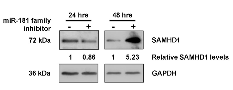
<strong>Figure 9 Regulated Expression Validation of SAMHD1 in MT2 CD4+ T-cells from leukemia patients
(Kohnken et al.,  2017)</strong><br>
SAMHD1 protein expression detected by anti-SAMHD1 antibodies (8007) was significantly increased by 5-fold at 48hr post-nucleofection with miR-181family inhibitor treatment.

<strong>Figure 1 Western Blot Validation in Human Daudi Cell Lines</strong><br>
Loading: 15 μg of lysates per lane.
Antibodies: SAMHD1 8007, 1 μg/mL, in (A: the absence and B: the presence of blocking peptide), (1h incubation at RT in 5% NFDM/TBST.
Secondary: Goat anti-rabbit IgG HRP conjugate at 1:10000 dilution.
<strong>Figure 2 Overexpression Validation in 293T Transfected Cells</strong><br>
Loading: 15 μg of lysates per lane.
Antibodies: SAMHD1 8007 (0.1 μg/mL), (1h incubation at RT in 5% NFDM/TBST.
Secondary: Goat anti-rabbit IgG HRP conjugate at 1:10000 dilution.
293 cells were transfected with (1) wild-type SAMHD1 or (2) SAMHD1 (mutation T592A).
<strong>Figure 3 Immunohistochemistry Validation of SAMHD1 in Human Brain Tissue </strong><br> 
Immunohistochemical analysis of paraffin-embedded Human Brain Tissue using anti- SAMHD1 antibody (8007) at 5 μg/ml. Tissue was fixed with formaldehyde and blocked with 10% serum for 1 h at RT; antigen retrieval was by heat mediation with a citrate buffer (pH6). Samples were incubated with primary antibody overnight at 4 ˚C. A goat anti-rabbit IgG H&L (HRP) at 1/250 was used as secondary. Counter stained with Hematoxylin.
<strong>Figure 4 Immunofluorescence Validation of SAMHD1 in Human Daudi cells</strong><br>
Immunofluorescent analysis of 4% paraformaldehyde-fixed Human Daudi Cells labeling SAMHD1 with 8007 at 20 μg/mL, followed by goat anti-rabbit IgG secondary antibody at 1/500 dilution (red).
<strong>Figure 5  KO Validation of SAMHD1 in xenograft mice (Kodigepalli et al., 2018)</strong><br> 
 THP-1 control and SAMHD1 KO (THP-1/KO) cells were injected into NSG (non-obese diabetic/severe combined
immune deficient-gamma) mice.  Protein expression levels of SAMHD1 were examined by Western blot with anti-SAMHD1 antibodies (8007) and SAMHD1 was not detected in THP-1/KO cells.
<strong>Figure 6 Overexpression Validation of SAMHD1 in CD4+ T-cells from a healthy donor and transformed CD4+ T-cell lines (Kohnken et al.,  2017) </strong><br>
MT1, MT2, SLB-1, and C8166 were from leukemia patients and HH, HuT78, and HuT102  were from cutaneous T-cell lymphoma (CTCL) patients.  SAMHD1 protein expression detected by anti-SAMHD1 antibodies (8007) was significantly increased in normal CD4+ T-cells as compared to leukemia- and CTCL- derived CD4+ T-cell lines. This blot is representative from four independent experiments with four healthy donors.
<strong>Figure 7 Overexpression  of SAMHD1 in CD4+ T-cells from Healthy Donors and S?zary Syndrome (SS) patients
(Kohnken et al.,  2017)</strong><br>
SAMHD1 protein expression detected by anti-SAMHD1 antibodies (8007) was significantly reduced in CD+ T-cells from 15 SS patients as compared to those from 7 healthy donors.
<strong>Figure 8 Regulated Expression Validation of SAMHD1 in CD4+ T-cells from a healthy donor (Kohnken et al.,  2017)</strong><br>
SAMHD1 protein expression detected by anti-SAMHD1 antibodies (8007) was significantly decreased by about 40% relative to control cells at 48hr post-nucleofection with miR-181b.
<strong>Figure 9 Regulated Expression Validation of SAMHD1 in MT2 CD4+ T-cells from leukemia patients
(Kohnken et al.,  2017)</strong><br>
SAMHD1 protein expression detected by anti-SAMHD1 antibodies (8007) was significantly increased by 5-fold at 48hr post-nucleofection with miR-181family inhibitor treatment.

Further Information

Additional Names:
SAM domain and HD domain 1, DCIP, CHBL2, HDDC1, MOP-5, SBBI88
Application Note:
WB: 1 μg/mL; IHC: 5 μg/mL; IF: 20 μg/mL.

Antibody validated: Western Blot, Immunohistochemistry, Immunofluorescence in human samples. All other applications and species not yet tested.
Background:
The SAM domain and HD domain 1 (SAMHD1) protein is upregulated in response to viral infection and is thought to play a role in innate immunity (1). SAMHD1 blocks the infection of HIV-1 and SIVdeltaVpx before reverse transcription in macrophages and dendritic cells (2), and this restriction is regulated by phosphorylation of SAMHD1 (3). Mutations in this gene have been associated with Aicardi-Goutieres syndrome (1).
Background References:
  • Rice et al. Nat. Genet. 2009; 41:829-32.
  • Hrecka et al. Nature 2011; 474:654-7.
  • Welbourn et al. J. Virol. 2013; 87:11516-24.
Buffer:
SAMHD1 antibody is supplied in PBS containing 0.02% sodium azide.
Concentration:
1 mg/mL
Conjugate:
Unconjugated
DISCLAIMER:
Optimal dilutions/concentrations should be determined by the end user. The information provided is a guideline for product use. This product is for research use only.
Immunogen:
Anti-SAMHD1 antibody (8007) was raised against a peptide corresponding to 18 amino acids near the carboxy terminus of human SAMHD1.
ISOFORMS:
Human SAMHD1 has 3 isoforms, including isoform 1 (626aa, 72kD), isoform 3 (556aa, 64kD), isoform 4 (591aa, 68kD). Mouse SAMHD1 has 2 isoforms, including (658aa, 76kD) and (651aa, 75kD). Rat SAMHD1 has only one isoform (620aa, 72kD). 8007 can detect human, mouse and rat.
NCBI Gene ID #:
25939
NCBI Official Name:
SAM domain and HD domain 1
NCBI Official Symbol:
SAMHD1
NCBI Organism:
homo sapiens
Physical State:
Liquid
PREDICTED MOLECULAR WEIGHT:
Predicted: 72kD

Observed: 72 kD
Protein Accession #:
NP_056289
Protein GI Number:
38016914
Purification:
SAMHD1 antibody is affinity chromatography purified via peptide column.
Research Area:
Innate Immunity
SPECIFICITY:
SAMHD1 antibody is human and mouse reactive.
Swissprot #:
Q9Y3Z3
User NOte:
Optimal dilutions for each application to be determined by the researcher.
VALIDATION:

KO Validation (Figure 5) shows SAMHD1 expression detected by anti-SAMHD1 antibodies (8007) was disrupted in multiple cells of SAMHD1 KO mice (figure 6).

Overexpression validation (Figure 2,6,7): SAMHD1 overexpression detected by anit-SAMHD1 antibodies (8007) was observed in normal cell lines stably across all healthy donors.

Regulated expression validation (Figure 8 & 9): SAMHD1 expression detected by anit-SAMHD1 antibodies (8007) was down-regulated by nucleofection with miR-181b at 48hr (figure 8), but up-regulated by  treatment of miR-181family inhibitor (figure 9).

References

  1. Kodigepalli et al. SAMHD1 modulates in vitro proliferation of acute myeloid leukemia-derived THP-1 cells through the PI3K-Akt-p27 axis. Cell Cycle. 2018;17(9):1124-1137. PMID: 29911928
  2. Kohnken et al. MicroRNA-181 contributes to downregulation of SAMHD1 expression in CD4+ T-cells derived from S?zary syndrome patients. Leuk Res. 2017;52:58-66. PMID: 27889686
  3. St Gelais et al. A Cyclin-Binding Motif in Human SAMHD1 Is Required for Its HIV-1 Restriction, dNTPase Activity, Tetramer Formation, and Efficient Phosphorylation. J Virol. 2018;92(6). PMID: 29321329
  4. Wang et al. Phosphorylation of mouse SAMHD1 regulates its restriction of human immunodeficiency virus type 1 infection, but not murine leukemia virus infection. Virology. 2016;487:273-84.PMID: 26580513
  5. Rebecca Kohnken. MicroRNAs in Cutaneous T-cell Lymphoma Pathogenesis. PhD thesis. The Ohio State University. 2017.PMID: