PPARGC1A Antibody

ProSci
Product Code: PSI-7793
Product Group: Primary Antibodies
Supplier: ProSci
CodeSizePrice
PSI-7793-0.02mg0.02mg£150.00
Quantity:
PSI-7793-0.1mg0.1mg£449.00
Quantity:
Prices exclude any Taxes / VAT

Overview

Host Type: Rabbit
Antibody Isotype: IgG
Antibody Clonality: Polyclonal
Antibody Clone: 7019499
Regulatory Status: RUO
Target Species:
  • Human
  • Mouse
  • Rat
Applications:
  • Enzyme-Linked Immunosorbent Assay (ELISA)
  • Western Blot (WB)
Storage:
PPARGC1A antibody can be stored at 4˚C for three months and -20˚C, stable for up to one year.

Images

1 / 10
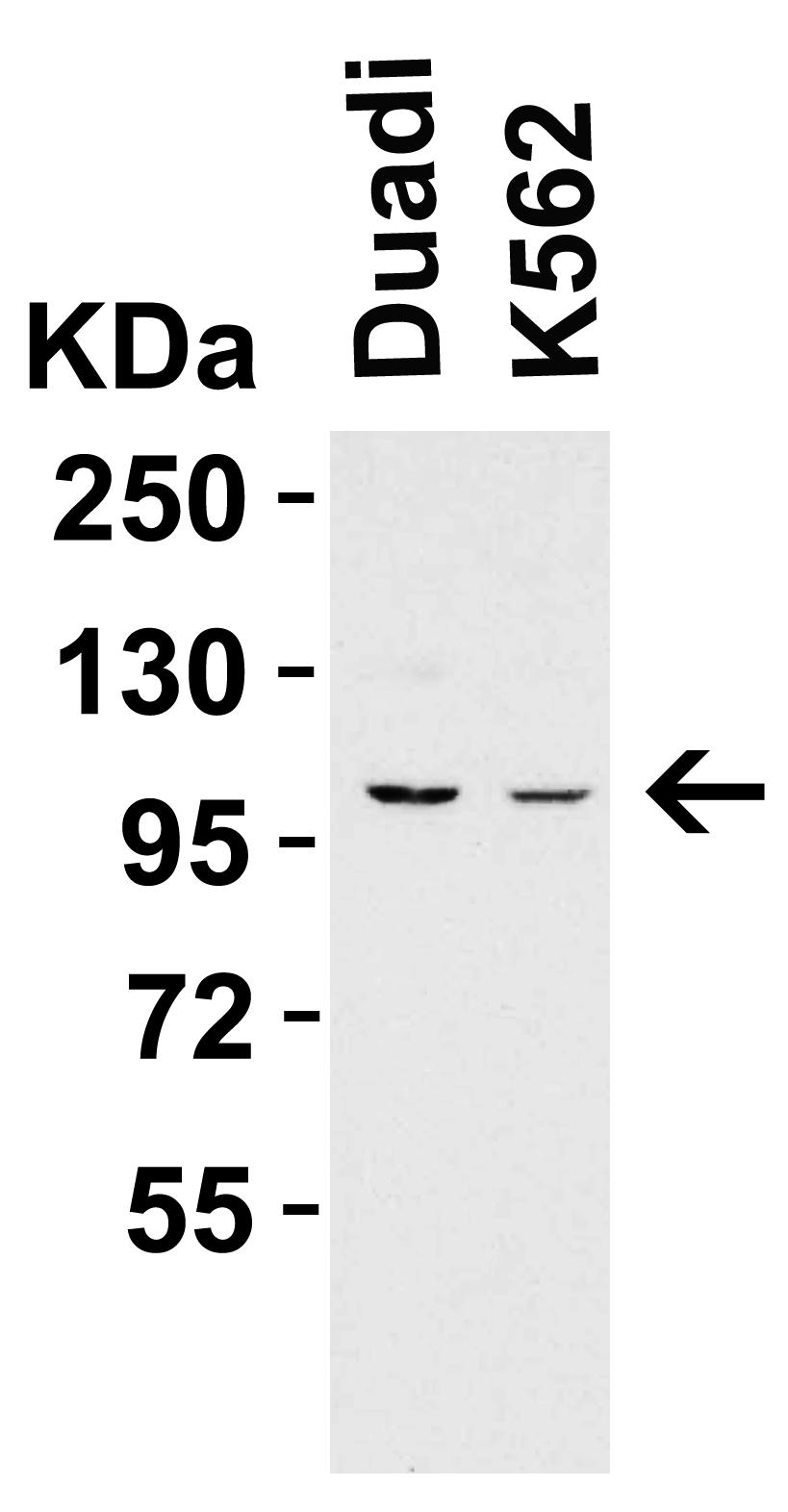
<strong>Figure 1 WB Validation  in Human Cell Lines </strong><br>
Loading: 15 μg of human cell lysate 
Antibodies:  PPARGC1A 7793, 2 μg/mL , 1 h incubation at RT in 5% NFDM/TBST. 
Secondary: Goat Anti-Rabbit IgG HRP conjugate at 1:10000 dilution.
2 / 10
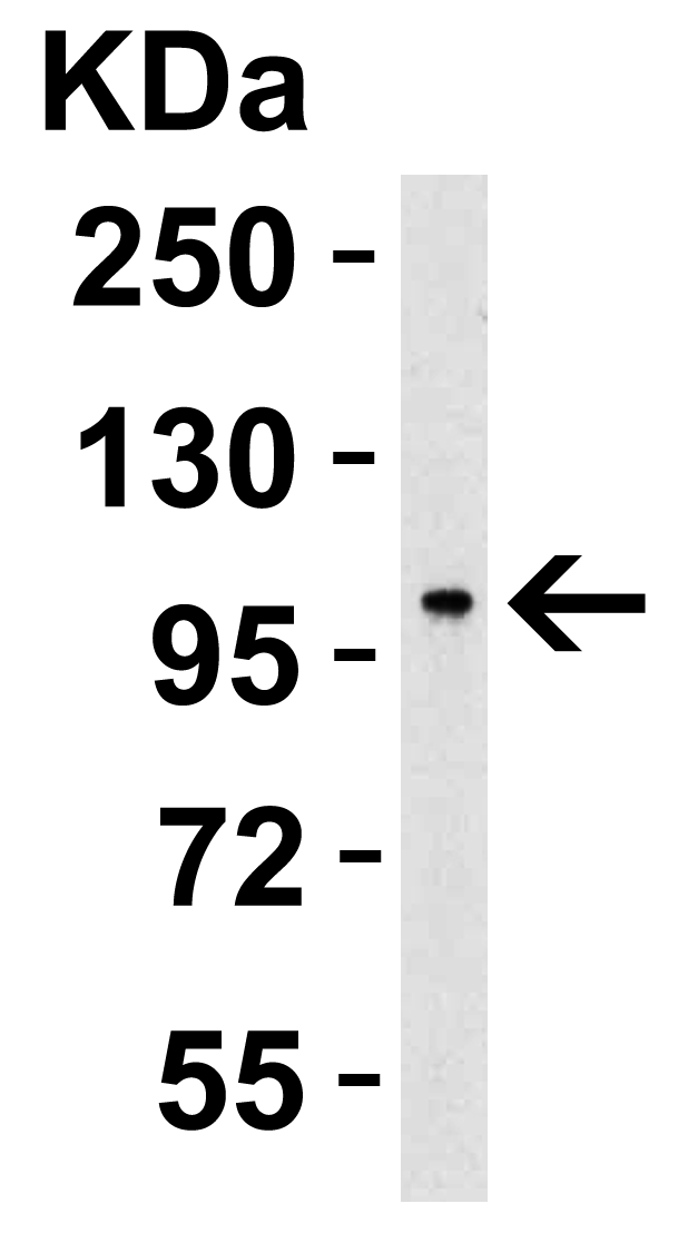
<strong>WB Validation  in Human Lung </strong><br>
Loading: 15 μg of human lung lysate 
Antibodies:  PPARGC1A 7793, 2 μg/mL , 1 h incubation at RT in 5% NFDM/TBST. 
Secondary: Goat Anti-Rabbit IgG HRP conjugate at 1:10000 dilution.
3 / 10
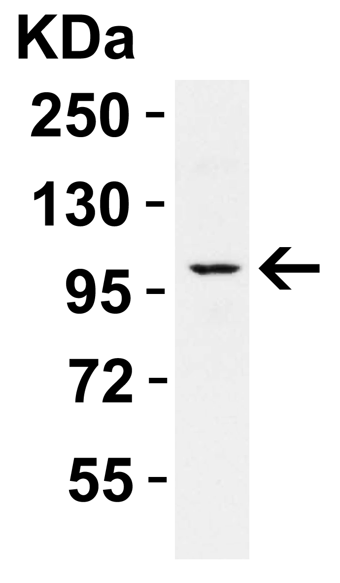
<strong>Figure 3 WB Validation  in Mouse Skin </strong><br>
Loading: 15 μg of mouse skin lysate 
Antibodies:  PPARGC1A 7793, 2 μg/mL , 1 h incubation at RT in 5% NFDM/TBST. 
Secondary: Goat Anti-Rabbit IgG HRP conjugate at 1:10000 dilution.
4 / 10
<strong>Figure 4  WB Validation  in Rat Kidney </strong><br>
Loading: 15 μg of rat kidney lysate 
Antibodies:  PPARGC1A 7793, 2 μg/mL , 1 h incubation at RT in 5% NFDM/TBST. 
Secondary: Goat Anti-Rabbit IgG HRP conjugate at 1:10000 dilution.
5 / 10
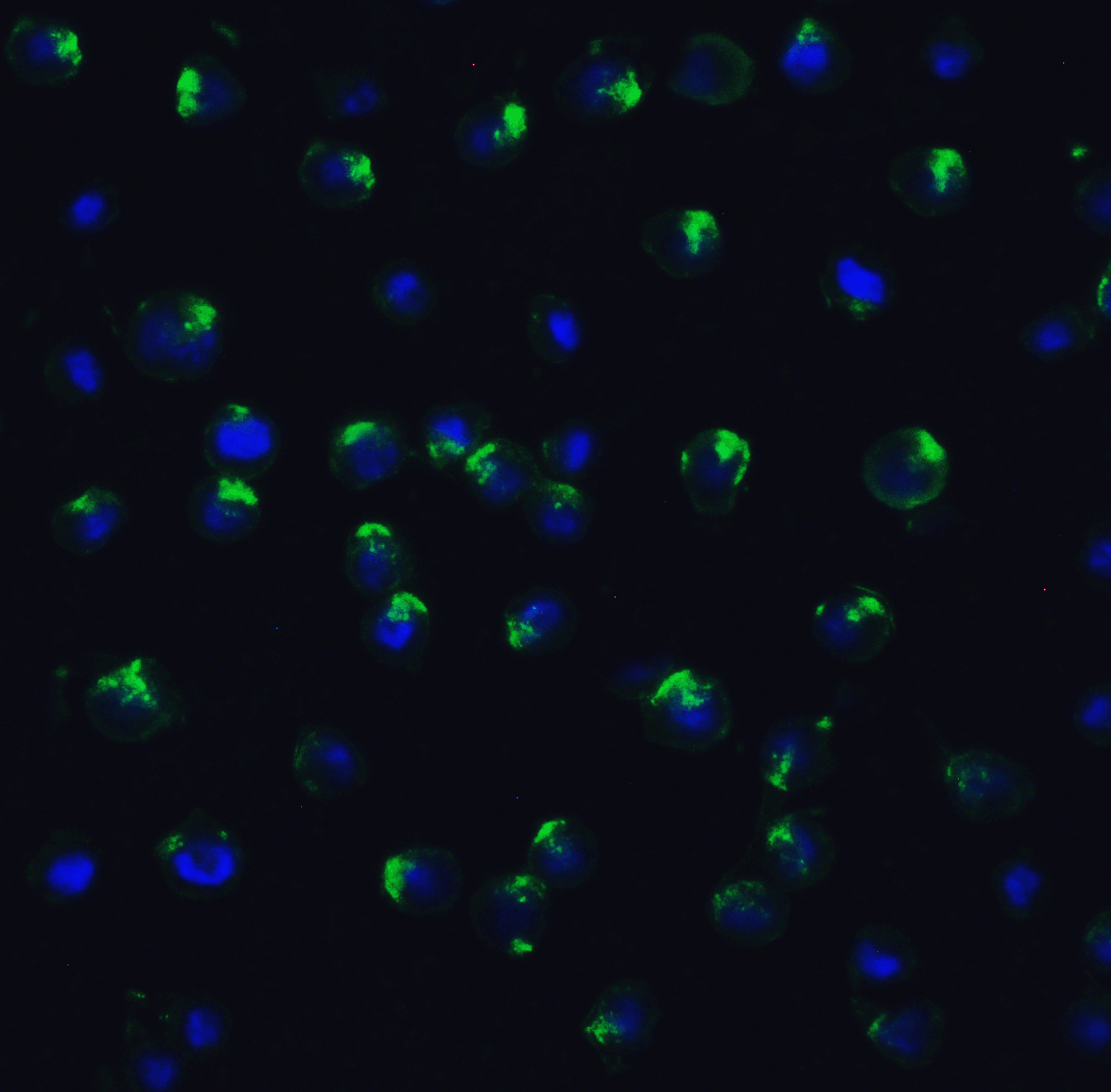
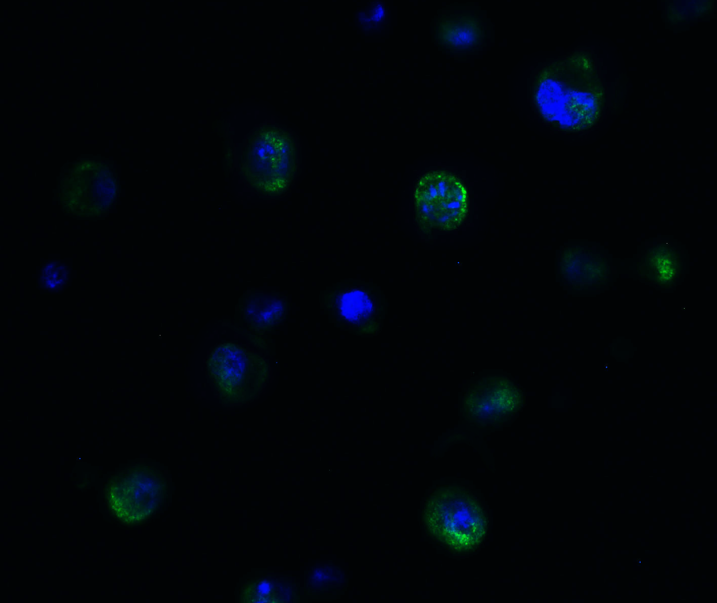
<strong>Figure 5 Immunofluorescence Validation of PPARGC1A in MCF7 Cells</strong><br>
Immunofluorescent analysis of 4% paraformaldehyde-fixed MCF7 cells labeling PPARGC1A with 7793 at 20 μg/mL, followed by goat anti-rabbit IgG secondary antibody at 1/500 dilution (green) and DAPI staining (blue).
6 / 10
<strong>Figure 6 Immunofluorescence Validation of PPARGC1A in HELA Cells</strong><br>
Immunofluorescent analysis of 4% paraformaldehyde-fixed HeLa cells labeling PPARGC1A with 7793 at 20 μg/mL, followed by goat anti-rabbit IgG secondary antibody at 1/500 dilution (green) and DAPI staining (blue).
7 / 10
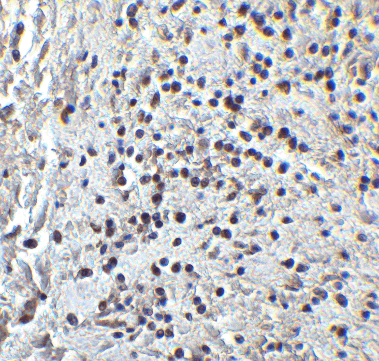
<strong>Figure 7  Immunohistochemistry Validation of PPARGC1A in Human Colon Carcinoma Tissue </strong><br> 
Immunohistochemical analysis of paraffin-embedded human colon carcinoma tissue using anti-PPARGC1A antibody (7793) at 1 μg/ml. Tissue was fixed with formaldehyde and blocked with 10% serum for 1 h at RT; antigen retrieval was by heat mediation with a citrate buffer (pH6). Samples were incubated with primary antibody overnight at 4˚C. A goat anti-rabbit IgG H&L (HRP) at 1/250 was used as secondary. Counter stained with Hematoxylin.
8 / 10
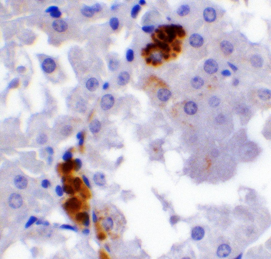
<strong>Figure 8  Immunohistochemistry Validation of PPARGC1A in Human Pancreas Tissue </strong><br> 
Immunohistochemical analysis of paraffin-embedded human pancreas tissue using anti-PPARGC1A antibody (7793) at 2 μg/ml. Tissue was fixed with formaldehyde and blocked with 10% serum for 1 h at RT; antigen retrieval was by heat mediation with a citrate buffer (pH6). Samples were incubated with primary antibody overnight at 4˚C. A goat anti-rabbit IgG H&L (HRP) at 1/250 was used as secondary. Counter stained with Hematoxylin.
9 / 10
<strong>Figure 9  Immunohistochemistry Validation of PPARGC1A in Mouse Stomach Tissue  </strong><br>
Immunohistochemical analysis of paraffin-embedded mouse stomach tissue using anti-PPARGC1A antibody (7793) at 2 μg/ml. Tissue was fixed with formaldehyde and blocked with 10% serum for 1 h at RT; antigen retrieval was by heat mediation with a citrate buffer (pH6). Samples were incubated with primary antibody overnight at 4˚C. A goat anti-rabbit IgG H&L (HRP) at 1/250 was used as secondary. Counter stained with Hematoxylin.
10 / 10
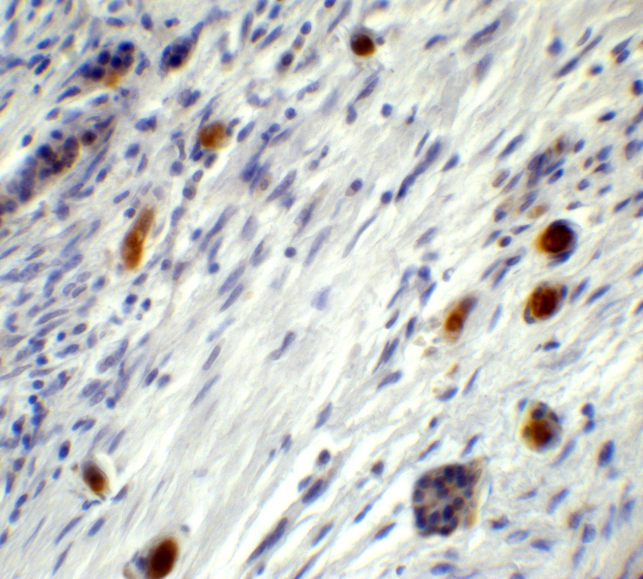
<strong>Figure 10  Immunohistochemistry Validation of PPARGC1A in Rat Kidney Tissue </strong><br> 
Immunohistochemical analysis of paraffin-embedded rat kidney tissue using anti-PPARGC1A antibody (7793) at 2 μg/ml. Tissue was fixed with formaldehyde and blocked with 10% serum for 1 h at RT; antigen retrieval was by heat mediation with a citrate buffer (pH6). Samples were incubated with primary antibody overnight at 4˚C. A goat anti-rabbit IgG H&L (HRP) at 1/250 was used as secondary. Counter stained with Hematoxylin.

<strong>Figure 1 WB Validation  in Human Cell Lines </strong><br>
Loading: 15 μg of human cell lysate 
Antibodies:  PPARGC1A 7793, 2 μg/mL , 1 h incubation at RT in 5% NFDM/TBST. 
Secondary: Goat Anti-Rabbit IgG HRP conjugate at 1:10000 dilution.
<strong>WB Validation  in Human Lung </strong><br>
Loading: 15 μg of human lung lysate 
Antibodies:  PPARGC1A 7793, 2 μg/mL , 1 h incubation at RT in 5% NFDM/TBST. 
Secondary: Goat Anti-Rabbit IgG HRP conjugate at 1:10000 dilution.
<strong>Figure 3 WB Validation  in Mouse Skin </strong><br>
Loading: 15 μg of mouse skin lysate 
Antibodies:  PPARGC1A 7793, 2 μg/mL , 1 h incubation at RT in 5% NFDM/TBST. 
Secondary: Goat Anti-Rabbit IgG HRP conjugate at 1:10000 dilution.
<strong>Figure 4  WB Validation  in Rat Kidney </strong><br>
Loading: 15 μg of rat kidney lysate 
Antibodies:  PPARGC1A 7793, 2 μg/mL , 1 h incubation at RT in 5% NFDM/TBST. 
Secondary: Goat Anti-Rabbit IgG HRP conjugate at 1:10000 dilution.
<strong>Figure 5 Immunofluorescence Validation of PPARGC1A in MCF7 Cells</strong><br>
Immunofluorescent analysis of 4% paraformaldehyde-fixed MCF7 cells labeling PPARGC1A with 7793 at 20 μg/mL, followed by goat anti-rabbit IgG secondary antibody at 1/500 dilution (green) and DAPI staining (blue).
<strong>Figure 6 Immunofluorescence Validation of PPARGC1A in HELA Cells</strong><br>
Immunofluorescent analysis of 4% paraformaldehyde-fixed HeLa cells labeling PPARGC1A with 7793 at 20 μg/mL, followed by goat anti-rabbit IgG secondary antibody at 1/500 dilution (green) and DAPI staining (blue).
<strong>Figure 7  Immunohistochemistry Validation of PPARGC1A in Human Colon Carcinoma Tissue </strong><br> 
Immunohistochemical analysis of paraffin-embedded human colon carcinoma tissue using anti-PPARGC1A antibody (7793) at 1 μg/ml. Tissue was fixed with formaldehyde and blocked with 10% serum for 1 h at RT; antigen retrieval was by heat mediation with a citrate buffer (pH6). Samples were incubated with primary antibody overnight at 4˚C. A goat anti-rabbit IgG H&L (HRP) at 1/250 was used as secondary. Counter stained with Hematoxylin.
<strong>Figure 8  Immunohistochemistry Validation of PPARGC1A in Human Pancreas Tissue </strong><br> 
Immunohistochemical analysis of paraffin-embedded human pancreas tissue using anti-PPARGC1A antibody (7793) at 2 μg/ml. Tissue was fixed with formaldehyde and blocked with 10% serum for 1 h at RT; antigen retrieval was by heat mediation with a citrate buffer (pH6). Samples were incubated with primary antibody overnight at 4˚C. A goat anti-rabbit IgG H&L (HRP) at 1/250 was used as secondary. Counter stained with Hematoxylin.
<strong>Figure 9  Immunohistochemistry Validation of PPARGC1A in Mouse Stomach Tissue  </strong><br>
Immunohistochemical analysis of paraffin-embedded mouse stomach tissue using anti-PPARGC1A antibody (7793) at 2 μg/ml. Tissue was fixed with formaldehyde and blocked with 10% serum for 1 h at RT; antigen retrieval was by heat mediation with a citrate buffer (pH6). Samples were incubated with primary antibody overnight at 4˚C. A goat anti-rabbit IgG H&L (HRP) at 1/250 was used as secondary. Counter stained with Hematoxylin.
<strong>Figure 10  Immunohistochemistry Validation of PPARGC1A in Rat Kidney Tissue </strong><br> 
Immunohistochemical analysis of paraffin-embedded rat kidney tissue using anti-PPARGC1A antibody (7793) at 2 μg/ml. Tissue was fixed with formaldehyde and blocked with 10% serum for 1 h at RT; antigen retrieval was by heat mediation with a citrate buffer (pH6). Samples were incubated with primary antibody overnight at 4˚C. A goat anti-rabbit IgG H&L (HRP) at 1/250 was used as secondary. Counter stained with Hematoxylin.

Further Information

Additional Names:
PPARGC1A Antibody: LEM6, PGC1, PGC1A, PGC-1v, PPARGC1, PGC-1(alpha), LEM6, Peroxisome proliferator-activated receptor gamma coactivator 1-alpha, Ligand effect modulator 6, PGC-1-alpha
Application Note:
WB: 2 μg/mL; IHC: 1-2 μg/mL; IF: 20 μg/mL.

Antibody validated: Western Blot in human, mouse and rat samples; Immunohistochemistry in human, mouse and rat samples; Immunofluorescence in human samples. All other applications and species not yet tested.
Background:
The peroxisome proliferator-activated receptor gamma coactivator 1-alpha (PPARGC1A), also known as LEM-6, is a transcriptional coactivator that regulates the genes involved in energy metabolism (1). PPARGC1A interacts with PPARgamma, which permits the interaction of PPARGC1A with multiple transcription factors. PPARGC1A can interact with, and regulate the activities of, cAMP response element binding protein (CREB) and nuclear respiratory factors (NRFs). It provides a direct link between external physiological stimuli and the regulation of mitochondrial biogenesis, and is a major factor that regulates muscle fiber type determination (2). PPARGC1A may be also involved in the development of obesity (3).
Background References:
  • Tsuemi T and La Spada AR. PGC-1a at the intersection of bioenergetics regulation and neuron function: from Huntington's disease to Parkinson's disease and beyond. Prog. Neurobiol. 2012; 97:142-51.
  • Kang C and Li Ji L. Role of PGC-1a signaling in skeletal muscle health and disease. Ann. NY Acad. Sci. 2012; 1271:110-7.
  • Liu C and Lin JD. PGC-1 coactivators in the control of energy metabolism. Acta Biochim. Biophys. Sin. 2011; 43:248-57.
Buffer:
PPARGC1A antibody is supplied in PBS containing 0.02% sodium azide.
Concentration:
1 mg/mL
Conjugate:
Unconjugated
DISCLAIMER:
Optimal dilutions/concentrations should be determined by the end user. The information provided is a guideline for product use. This product is for research use only.
Homology:
Predicted species reactivity based on immunogen sequence: Bovine: (100%), Pig: (94%)
Immunogen:
PPARGC1A antibody was raised against a 17 amino acid peptide near the amino terminus of human PPARGC1A.

The immunogen is located within amino acids 200 - 250 of PPARGC1A.
NCBI Gene ID #:
10891
NCBI Official Name:
peroxisome proliferator-activated receptor gamma, coactivator 1 alpha
NCBI Official Symbol:
PPARGC1A
NCBI Organism:
homo sapiens
Physical State:
Liquid
PREDICTED MOLECULAR WEIGHT:
Predicted: 91kD

Observed: 100 kD
Protein Accession #:
NP_037393
Protein GI Number:
7019499
Purification:
PPARGC1A antibody is affinity chromatography purified via peptide column.
Research Area:
Obesity
SPECIFICITY:
PPARGC1A antibody is human, mouse and rat reactive. At least three isoforms of PPARGC1A are known to exist; this antibody will detect all three isoforms.
Swissprot #:
Q9UBK2
User NOte:
Optimal dilutions for each application to be determined by the researcher.