VISTA Antibody [4C4]
| Code | Size | Price |
|---|
| PSI-RF16071-0.02mg | 0.02mg | £150.00 |
Quantity:
| PSI-RF16071-0.1mg | 0.1mg | £515.00 |
Quantity:
Prices exclude any Taxes / VAT
Overview
Host Type: Mouse
Antibody Isotype: IgG1
Antibody Clonality: Monoclonal
Antibody Clone: 4C4
Regulatory Status: RUO
Target Species: Human
Applications:
- Enzyme-Linked Immunosorbent Assay (ELISA)
- Flow Cytometry
- Immunocytochemistry (ICC)
- Immunofluorescence (IF)
- Immunohistochemistry- Paraffin Embedded (IHC-P)
- Western Blot (WB)
Shipping:
blue ice
Storage:
VISTA antibody can be stored at 4˚C for three months and -20˚C, stable for up to one year. As with all antibodies care should be taken to avoid repeated freeze thaw cycles. Antibodies should not be exposed to prolonged high temperatures.
Images
Documents
Further Information
Additional Names:
VISTA Antibody: VISTA molecule, VSIR, B7-H5, B7H5, GI24, PP2135, SISP1, DD1alpha, VISTA, C10orf54, chromosome 10 open reading frame 54, PD-1H, V-set immunoregulatory receptor, V-Type Immunoglobulin Domain-Containing Suppressor Of T-Cell Activation, Chromosome 10 Open Reading Frame 54
Application Note:
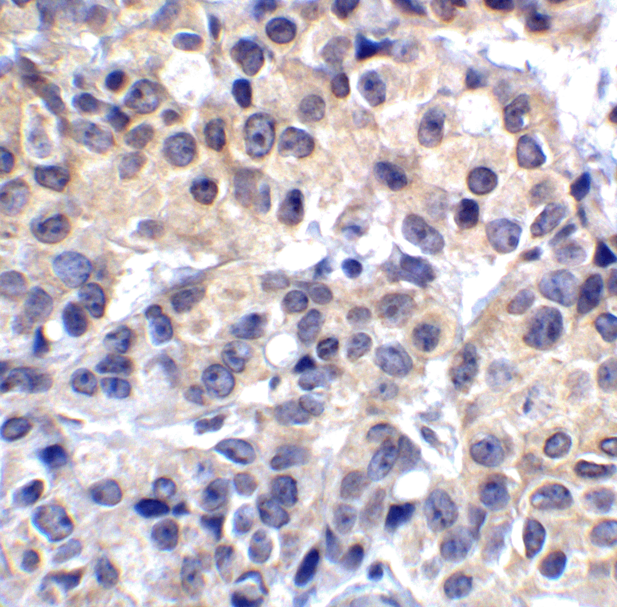
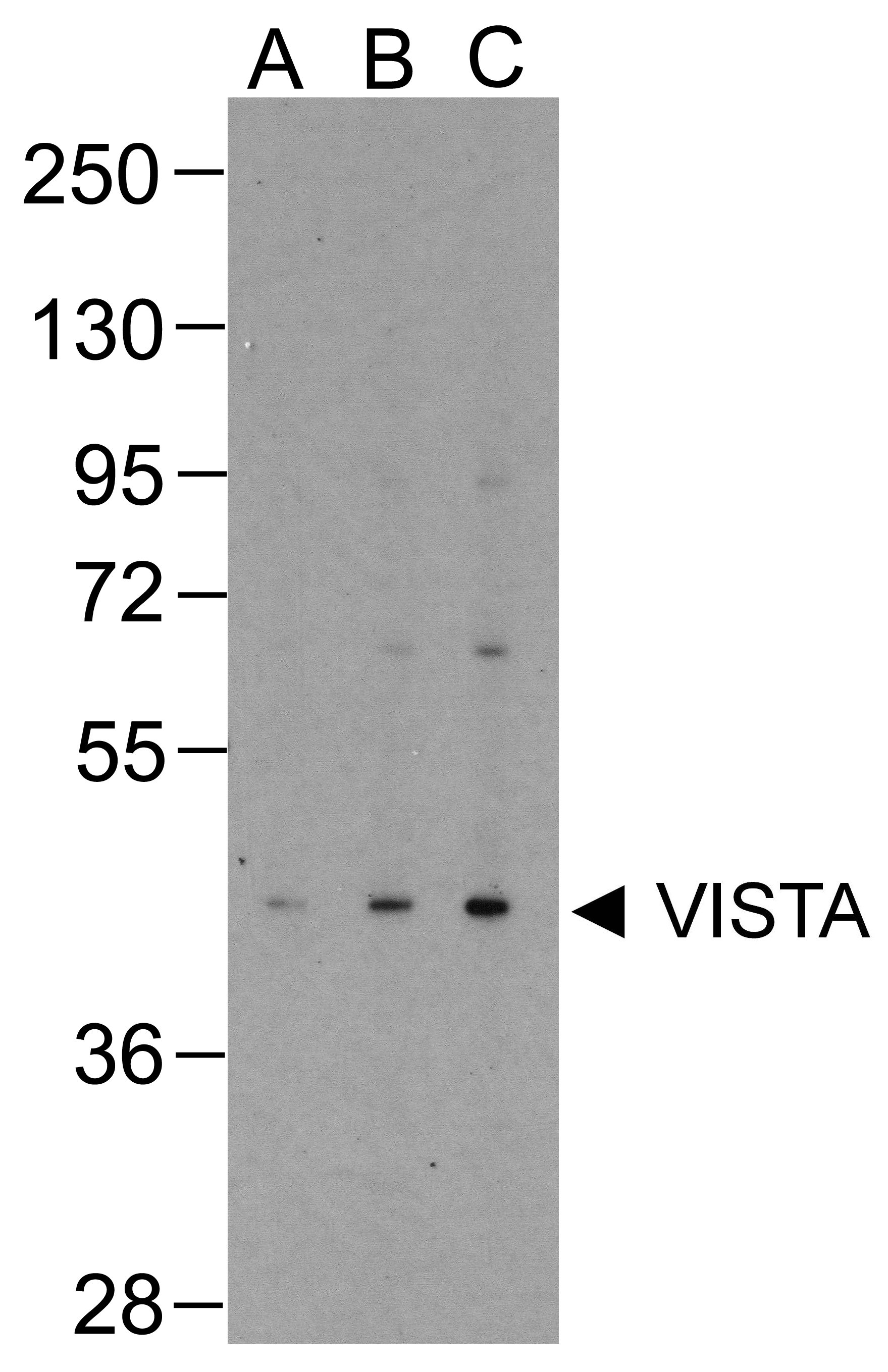
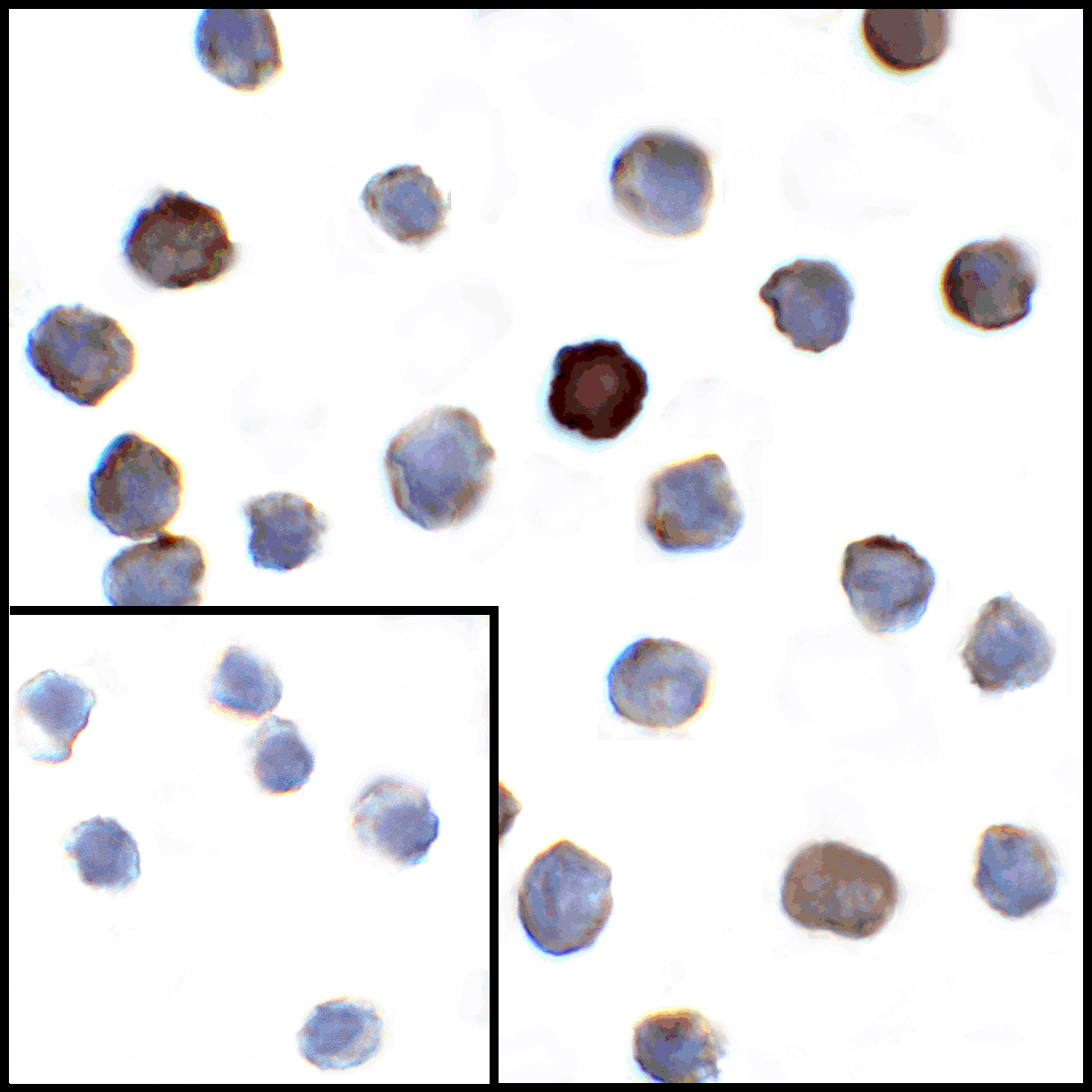
VISTA antibody can be used for detection of VISTA by Western blot at 0.25 - 1 μg/mL. Antibody can also be used for immunohistochemistry starting at 2 μg/mL and immunocytochemistry starting at 1 μg/mL. For immunofluorescence start at 2 μg/mL.
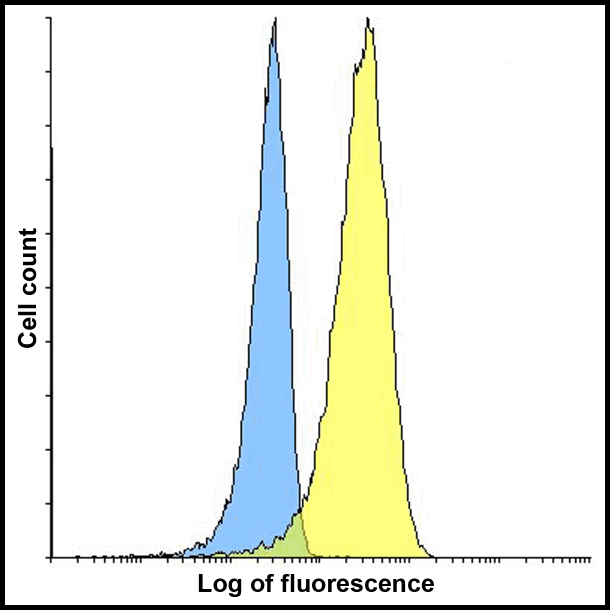
Antibody validated: Western Blot in human samples; Immunohistochemistry in human samples; Immunocytochemistry in human samples; Immunofluorescence in human samples and Flow Cytometry in mouse samples. All other applications and species not yet tested.
Antibody validated: Western Blot in human samples; Immunohistochemistry in human samples; Immunocytochemistry in human samples; Immunofluorescence in human samples and Flow Cytometry in mouse samples. All other applications and species not yet tested.
Background:
VISTA Antibody: VISTA/B7-H5/platelet receptor Gi24 is a single-pass type I membrane protein located at the cell surface. It is an immunoregulatory receptor which can inhibit T-cell response and may promote differentiation of embryonic stem cells, by inhibiting the BMP4 signaling pathway. The protein can be cleaved by MMP14, and stimulate MMP14-mediated MMP2 activation.
Background References:
- Mayya V., et al . Quantitative phosphoproteomic analysis of T cell receptor signaling reveals system-wide modulation of protein-protein interactions. 2009, Sci. Signal. 2:RA46-RA46.
- Sakr M.A., et al.,GI24 enhances tumor invasiveness by regulating cell surface membrane-type 1 matrix metalloproteinase. 2010, Cancer Sci. 101:2368-2374.
Buffer:
VISTA Antibody is supplied in PBS containing 0.02% sodium azide and 50% glycerol.
Concentration:
1 mg/mL
Conjugate:
Unconjugated
DISCLAIMER:
Optimal dilutions/concentrations should be determined by the end user. The information provided is a guideline for product use. This product is for research use only.
Immunogen:
VISTA antibody was raised against the extracellular domain of human VISTA.
NCBI Gene ID #:
64115
NCBI Official Name:
VISTA molecule
NCBI Official Symbol:
VSIR
NCBI Organism:
Homo sapiens
Physical State:
Liquid
PREDICTED MOLECULAR WEIGHT:
Predicted: 31 kDa
Observed: 47 kDa
Observed: 47 kDa
Protein Accession #:
NP_071436
Protein GI Number:
64115
Purification:
VISTA Antibody is supplied as protein A purified IgG1.
Research Area:
Immunology
Swissprot #:
Q9H7M9
User NOte:
Optimal dilutions for each application to be determined by the researcher.


![Immunofluorescence of VISTA in transfected HEK293 cells with VISTA antibody at 2 μg/mL. <br><br>Green: VISTA Antibody [4C4] (RF16071) <br> Blue: DAPI staining Immunofluorescence of VISTA in transfected HEK293 cells with VISTA antibody at 2 μg/mL. <br><br>Green: VISTA Antibody [4C4] (RF16071) <br> Blue: DAPI staining](https://www.prosci-inc.com/static-images/VISTA-Antibody-4C4_IF_RF16071.gif)
![Immunofluorescence of VISTA in human lymphoma tissue with VISTA antibody at 5 μg/mL. <br><br>Red: VISTA Antibody [4C4] (RF16071) <br> Blue: DAPI staining Immunofluorescence of VISTA in human lymphoma tissue with VISTA antibody at 5 μg/mL. <br><br>Red: VISTA Antibody [4C4] (RF16071) <br> Blue: DAPI staining](https://www.prosci-inc.com/static-images/VISTA-Antibody-4C4_IF-2_RF16071.gif)
![Immunofluorescence of VISTA in human spleen tissue with VISTA antibody at 10 μg/mL. <br><br>Red: VISTA Antibody [4C4] (RF16071) <br> Blue: DAPI staining Immunofluorescence of VISTA in human spleen tissue with VISTA antibody at 10 μg/mL. <br><br>Red: VISTA Antibody [4C4] (RF16071) <br> Blue: DAPI staining](https://www.prosci-inc.com/static-images/VISTA-Antibody-4C4_IF-3_RF16071.gif)
