PDL1 Antibody [6H10]

ProSci
Product Code: PSI-RF16035
Product Group: Primary Antibodies
Supplier: ProSci
CodeSizePrice
PSI-RF16035-0.02mg0.02mg£150.00
Quantity:
PSI-RF16035-0.1mg0.1mg£515.00
Quantity:
Prices exclude any Taxes / VAT

Overview

Host Type: Mouse
Antibody Isotype: IgG1
Antibody Clonality: Monoclonal
Antibody Clone: 6H10
Regulatory Status: RUO
Target Species: Human
Applications:
  • Enzyme-Linked Immunosorbent Assay (ELISA)
  • Immunocytochemistry (ICC)
  • Immunofluorescence (IF)
  • Immunohistochemistry- Paraffin Embedded (IHC-P)
  • Western Blot (WB)
Shipping:
blue ice
Storage:
PD-L1 antibody can be stored at 4˚C for three months and -20˚C, stable for up to one year. As with all antibodies care should be taken to avoid repeated freeze thaw cycles. Antibodies should not be exposed to prolonged high temperatures.

Images

1 / 10
<strong>Figure 1 Overexpression Validation of PD-L1 in 293 Cells </strong><br>
Loading: 15 μg of lysates per lane.
Antibodies: RF16035 (A, 0.25 μg/mL; B, 0.5 μg/mL), 1 h incubation at RT in 5% NFDM/TBST.
Secondary: Goat anti-mouse IgG HRP conjugate at 1:5000 dilution.
2 / 10
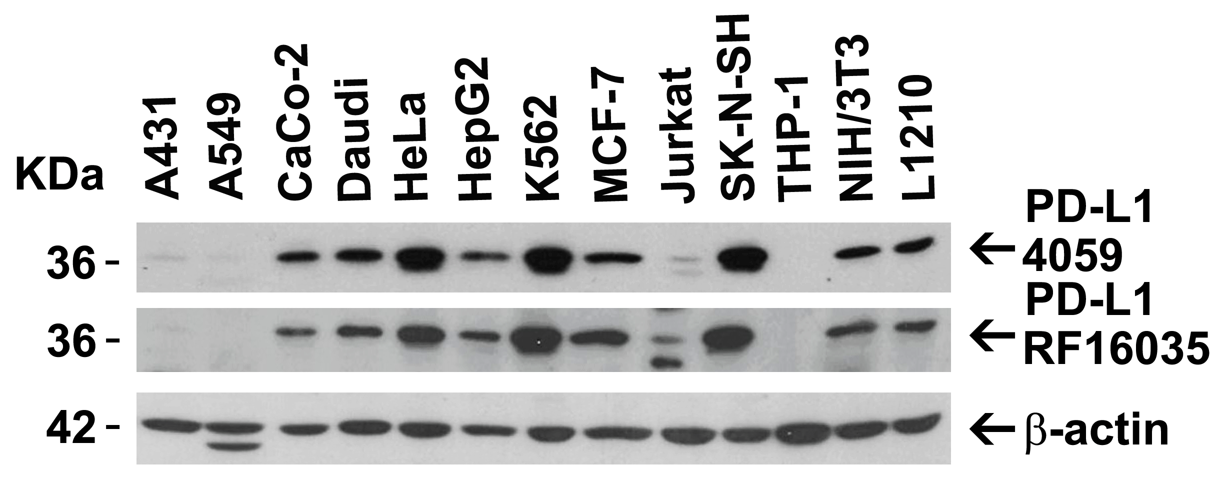
<strong>Figure 2 Independent Antibody Validation (IAV) via Protein Expression Profile in Cell Lines</strong><br>
Loading: 15 μg of lysates per lane.
Antibodies: 4059 (2 μg/mL), RF16035 (2 μg/mL), and beta-actin (1 μg/mL), 1 h incubation at RT in 5% NFDM/TBST.
Secondary: Goat anti-rabbit and or anti-mouse IgG HRP conjugate at 1:10000 and 1:5000 dilution, respectively.
3 / 10
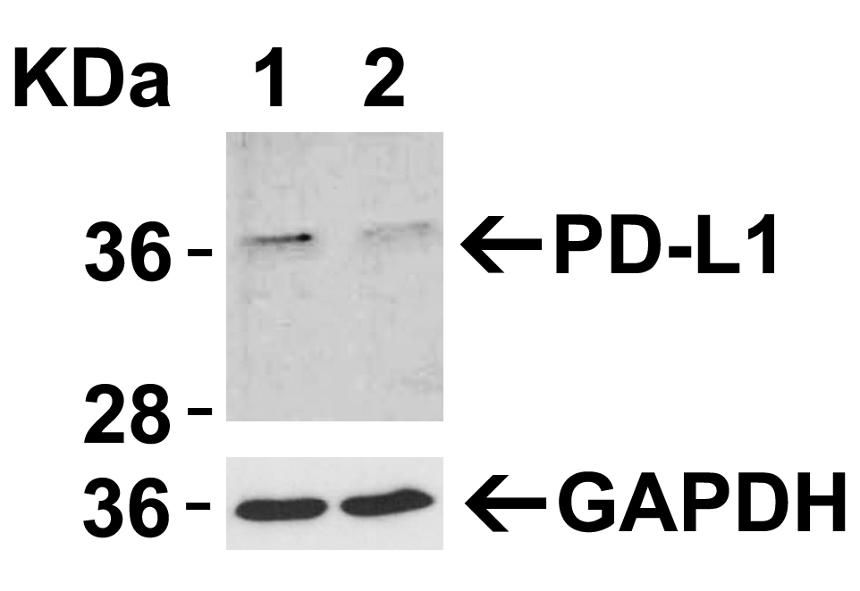
<strong>Figure 3 Validation with PD-L1 siRNA Knockdown in HeLa Cells</strong><br> 
HeLa cells were transfected with control siRNAs (lane 1) or PD-L1 siRNAs (lane 2) 
Loading: 10 μg of HeLa whole cell lysates per lane.
Antibodies: RF16035 (2 μg/mL) and GAPDH (3783, 0.02 μg/mL), 1 h incubation at RT in 5% NFDM/TBST.
Secondary: Goat anti-mouse IgG HRP conjugate at 1:5000 dilution.
4 / 10
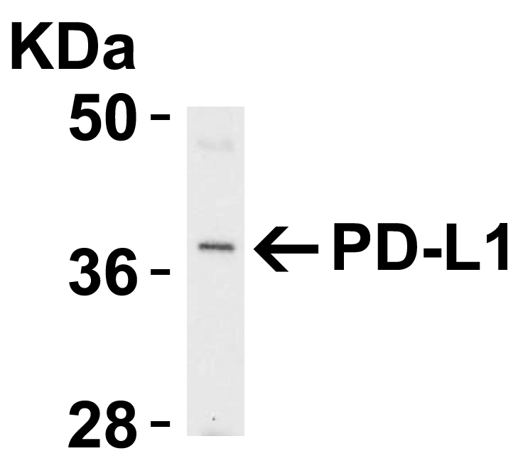
<strong>Figure 4 Western Blot Validation of PD-L1 in Raji Cells </strong><br> 
Loading: Lysates/proteins at 15 μg per lane.
Antibodies: RF16035 (4 μg/mL). 1 h incubation at RT in 5% NFDM/TBST.
Secondary: Goat anti-mouse IgG HRP conjugate at 1:10000 dilution.
5 / 10
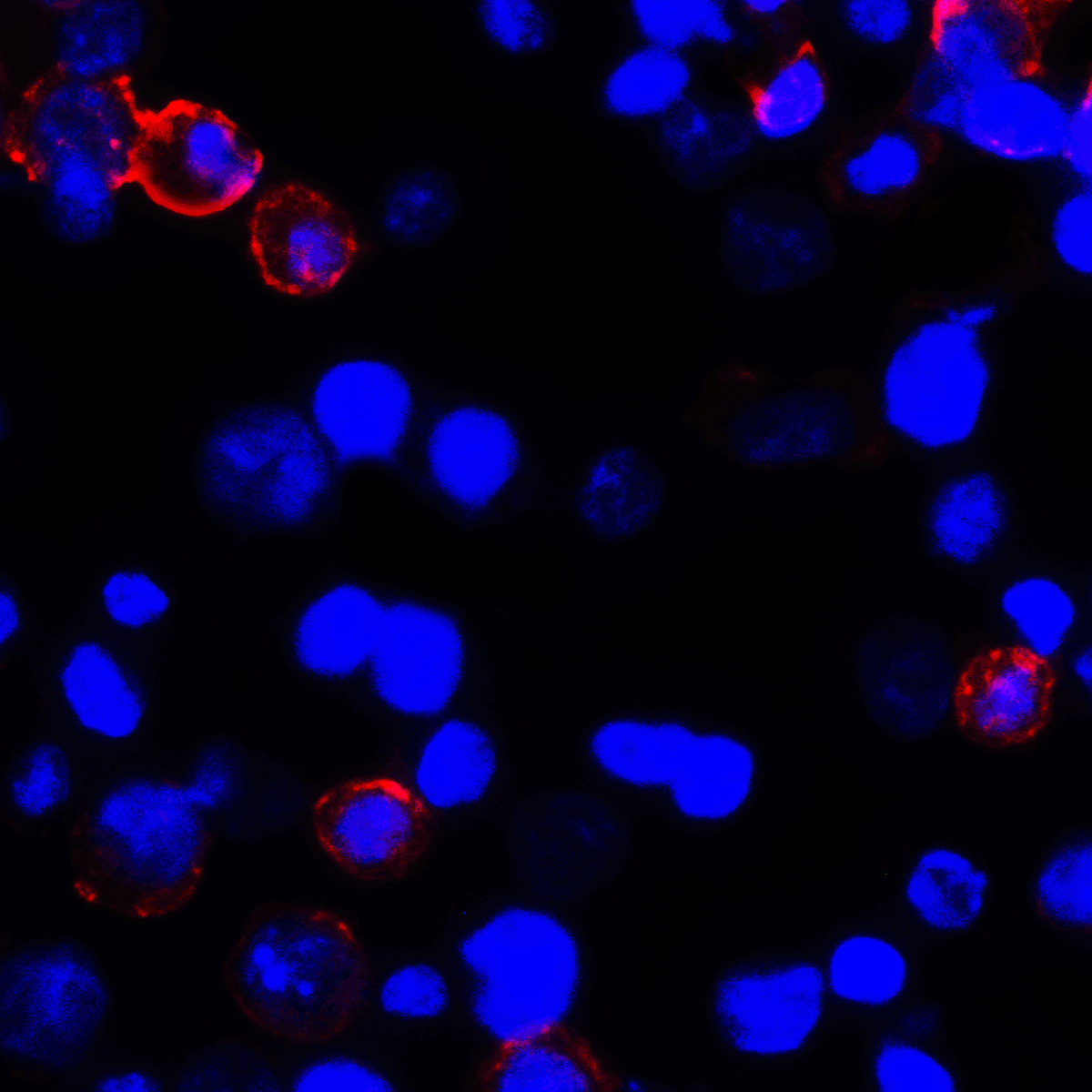
<strong>Figure 5 Immunofluorescence Validation of PD-L1 in Transfected 293 Cells</strong><br> 
Immunofluorescent analysis of 4% paraformaldehyde-fixed PD-L1 transfected 293 cells labeling PD-L1 with RF16035 at 2 μg/mL, followed by goat anti-mouse IgG secondary antibody at 1/500 dilution (red) and DAPI staining (blue).
6 / 10
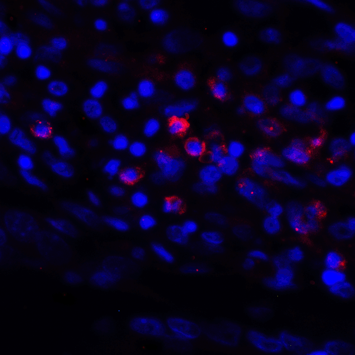
<strong>Figure 6 Immunofluorescence Validation of PD-L1 in Human Stomach Carcinoma Tissue</strong><br> 
Immunofluorescent analysis of 4% paraformaldehyde-fixed human stomach carcinoma tissue labeling PD-L1 with RF16035 at 2 μg/mL, followed by goat anti-mouse IgG secondary antibody at 1/500 dilution (red) and DAPI staining (blue).
7 / 10
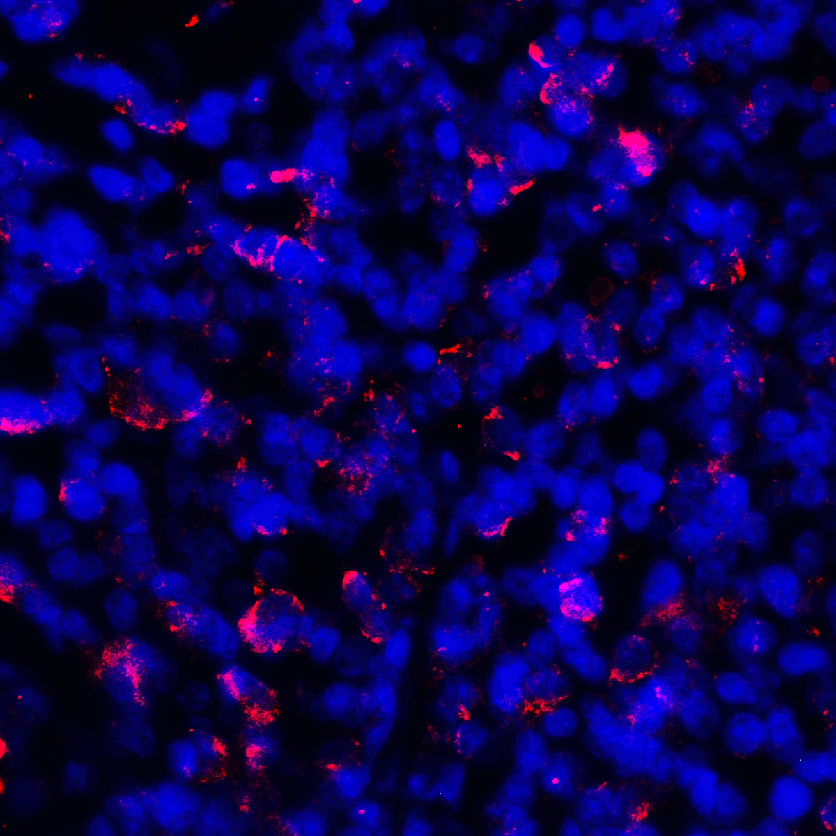
<strong>Figure 7 Immunofluorescence Validation of PD-L1 in Human Tonsil Tissue</strong><br> 
Immunofluorescent analysis of 4% paraformaldehyde-fixed human tonsil tissue labeling PD-L1 with RF16035 at 2 μg/mL, followed by goat anti-mouse IgG secondary antibody at 1/500 dilution (red) and DAPI staining (blue).
8 / 10
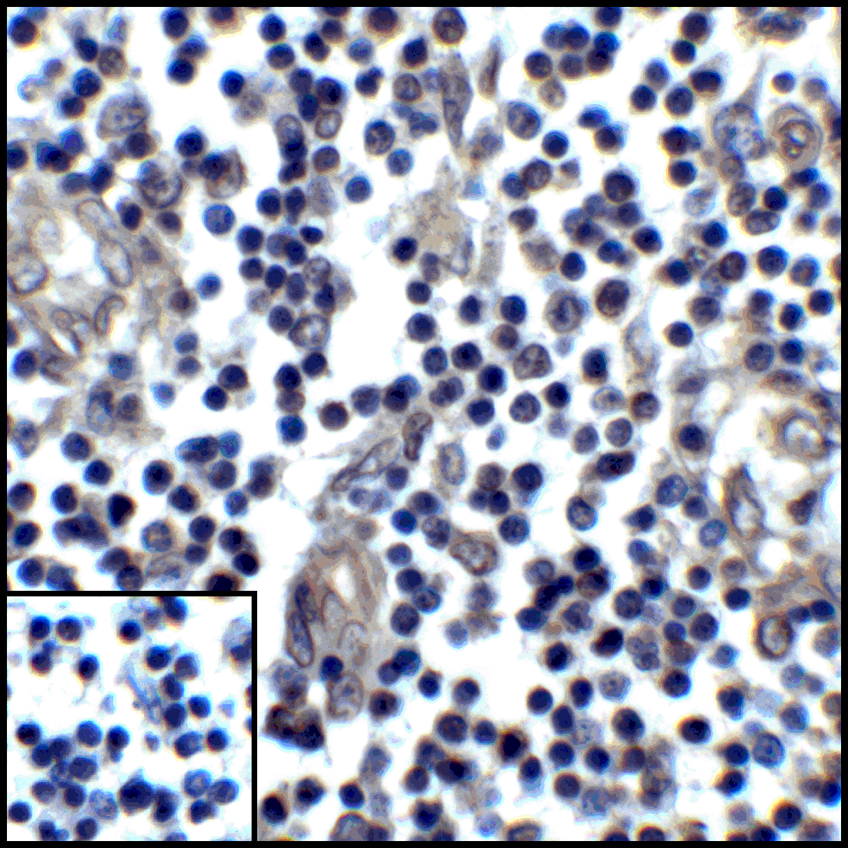
<strong>Figure 8 Immunohistochemistry Validation of PD-L1 in Human Stomach Carcinoma Tissue </strong><br>
Immunohistochemical analysis of paraffin-embedded human stomach carcinoma tissue using anti-PD-L1 antibody (RF16035) at 5 μg/ml. Tissue was fixed with formaldehyde and blocked with 10% serum for 1 h at RT; antigen retrieval was by heat mediation with a citrate buffer (pH6). Samples were incubated with primary antibody overnight at 4˚C. A goat anti-mouse IgG H&L (HRP) at 1/250 was used as secondary. Counter stained with Hematoxylin. Lower left: Use mouse IgG antibody at 1 μg/ml as control.
9 / 10
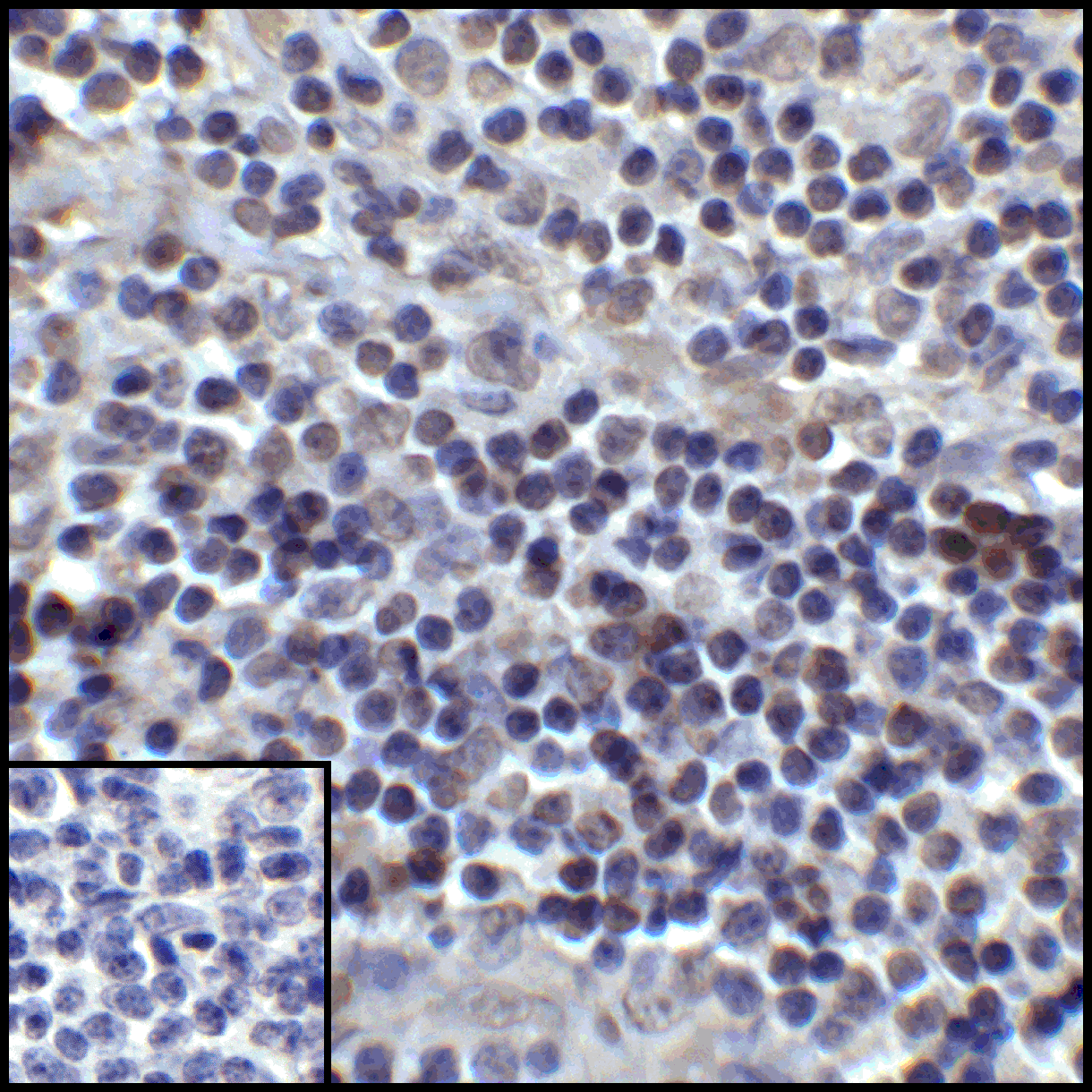
<strong>Figure 9 Immunohistochemistry Validation of PD-L1 in Human Tonsil Carcinoma Tissue </strong><br>
Immunohistochemical analysis of paraffin-embedded human tonsil tissue using anti-PD-L1 antibody (RF16035) at 5 μg/ml. Tissue was fixed with formaldehyde and blocked with 10% serum for 1 h at RT; antigen retrieval was by heat mediation with a citrate buffer (pH6). Samples were incubated with primary antibody overnight at 4˚C. A goat anti-mouse IgG H&L (HRP) at 1/250 was used as secondary. Counter stained with Hematoxylin. Lower left: Use mouse IgG antibody at 1 μg/ml as control.
10 / 10
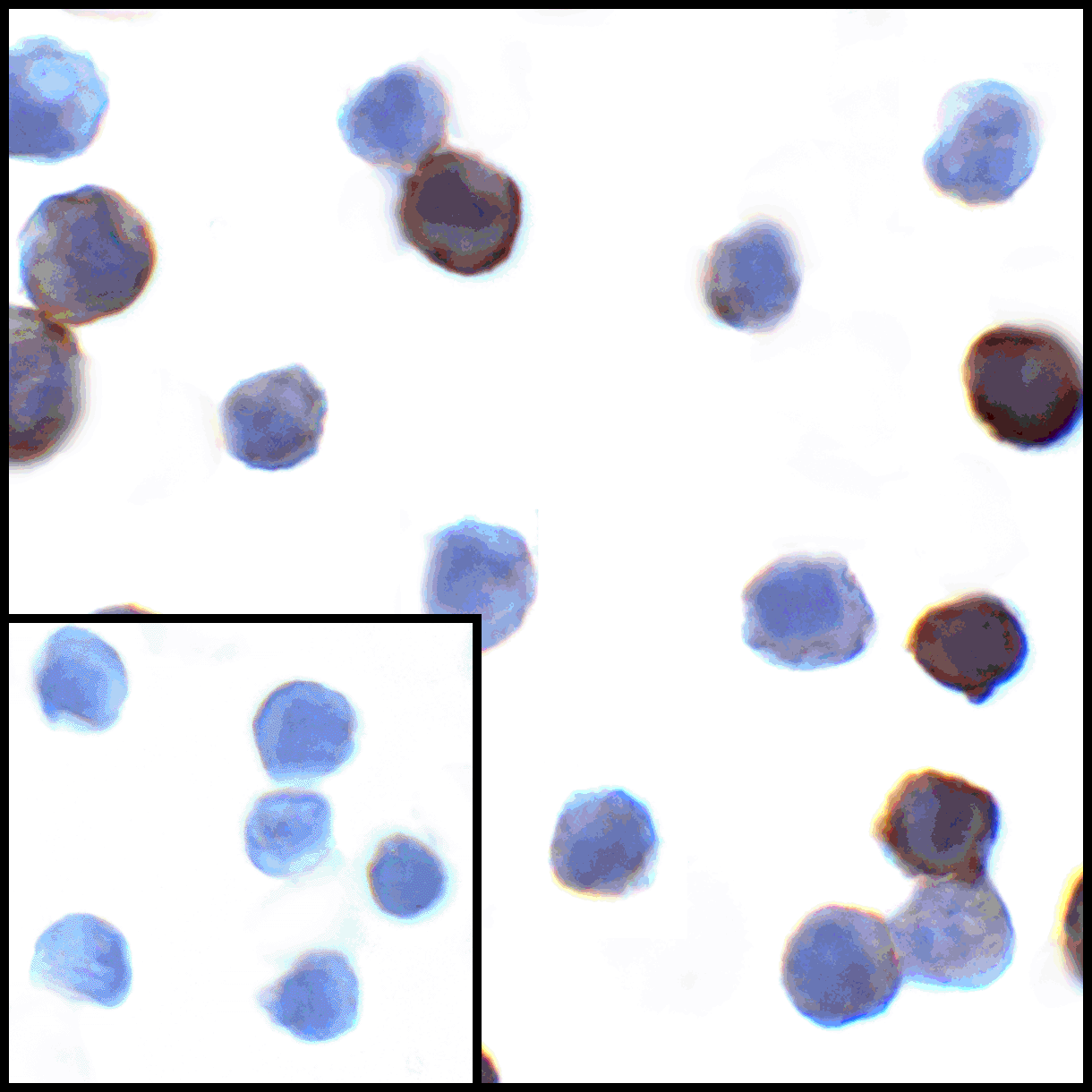
<strong> Figure 10 Immunocytochemistry Validation of PD-L1 in PD-L1 Overexpressed 293 Cells </strong><br>
Immunocytochemical analysis of PD-L1 transfected 293 cells using anti- PD-L1 antibody (RF16035) at 1 μg/mL. Cells was fixed with formaldehyde and blocked with 10% serum for 1 h at RT; antigen retrieval was by heat mediation with a citrate buffer (pH6). Samples were incubated with primary antibody overnight at 4˚ C. A goat anti-rabbit IgG H&L (HRP) at 1/250 was used as secondary. Counter stained with Hematoxylin.  Lower left: Use mouse IgG antibody at 1 μg/mL  as control.

<strong>Figure 1 Overexpression Validation of PD-L1 in 293 Cells </strong><br>
Loading: 15 μg of lysates per lane.
Antibodies: RF16035 (A, 0.25 μg/mL; B, 0.5 μg/mL), 1 h incubation at RT in 5% NFDM/TBST.
Secondary: Goat anti-mouse IgG HRP conjugate at 1:5000 dilution.
<strong>Figure 2 Independent Antibody Validation (IAV) via Protein Expression Profile in Cell Lines</strong><br>
Loading: 15 μg of lysates per lane.
Antibodies: 4059 (2 μg/mL), RF16035 (2 μg/mL), and beta-actin (1 μg/mL), 1 h incubation at RT in 5% NFDM/TBST.
Secondary: Goat anti-rabbit and or anti-mouse IgG HRP conjugate at 1:10000 and 1:5000 dilution, respectively.
<strong>Figure 3 Validation with PD-L1 siRNA Knockdown in HeLa Cells</strong><br> 
HeLa cells were transfected with control siRNAs (lane 1) or PD-L1 siRNAs (lane 2) 
Loading: 10 μg of HeLa whole cell lysates per lane.
Antibodies: RF16035 (2 μg/mL) and GAPDH (3783, 0.02 μg/mL), 1 h incubation at RT in 5% NFDM/TBST.
Secondary: Goat anti-mouse IgG HRP conjugate at 1:5000 dilution.
<strong>Figure 4 Western Blot Validation of PD-L1 in Raji Cells </strong><br> 
Loading: Lysates/proteins at 15 μg per lane.
Antibodies: RF16035 (4 μg/mL). 1 h incubation at RT in 5% NFDM/TBST.
Secondary: Goat anti-mouse IgG HRP conjugate at 1:10000 dilution.
<strong>Figure 5 Immunofluorescence Validation of PD-L1 in Transfected 293 Cells</strong><br> 
Immunofluorescent analysis of 4% paraformaldehyde-fixed PD-L1 transfected 293 cells labeling PD-L1 with RF16035 at 2 μg/mL, followed by goat anti-mouse IgG secondary antibody at 1/500 dilution (red) and DAPI staining (blue).
<strong>Figure 6 Immunofluorescence Validation of PD-L1 in Human Stomach Carcinoma Tissue</strong><br> 
Immunofluorescent analysis of 4% paraformaldehyde-fixed human stomach carcinoma tissue labeling PD-L1 with RF16035 at 2 μg/mL, followed by goat anti-mouse IgG secondary antibody at 1/500 dilution (red) and DAPI staining (blue).
<strong>Figure 7 Immunofluorescence Validation of PD-L1 in Human Tonsil Tissue</strong><br> 
Immunofluorescent analysis of 4% paraformaldehyde-fixed human tonsil tissue labeling PD-L1 with RF16035 at 2 μg/mL, followed by goat anti-mouse IgG secondary antibody at 1/500 dilution (red) and DAPI staining (blue).
<strong>Figure 8 Immunohistochemistry Validation of PD-L1 in Human Stomach Carcinoma Tissue </strong><br>
Immunohistochemical analysis of paraffin-embedded human stomach carcinoma tissue using anti-PD-L1 antibody (RF16035) at 5 μg/ml. Tissue was fixed with formaldehyde and blocked with 10% serum for 1 h at RT; antigen retrieval was by heat mediation with a citrate buffer (pH6). Samples were incubated with primary antibody overnight at 4˚C. A goat anti-mouse IgG H&L (HRP) at 1/250 was used as secondary. Counter stained with Hematoxylin. Lower left: Use mouse IgG antibody at 1 μg/ml as control.
<strong>Figure 9 Immunohistochemistry Validation of PD-L1 in Human Tonsil Carcinoma Tissue </strong><br>
Immunohistochemical analysis of paraffin-embedded human tonsil tissue using anti-PD-L1 antibody (RF16035) at 5 μg/ml. Tissue was fixed with formaldehyde and blocked with 10% serum for 1 h at RT; antigen retrieval was by heat mediation with a citrate buffer (pH6). Samples were incubated with primary antibody overnight at 4˚C. A goat anti-mouse IgG H&L (HRP) at 1/250 was used as secondary. Counter stained with Hematoxylin. Lower left: Use mouse IgG antibody at 1 μg/ml as control.
<strong> Figure 10 Immunocytochemistry Validation of PD-L1 in PD-L1 Overexpressed 293 Cells </strong><br>
Immunocytochemical analysis of PD-L1 transfected 293 cells using anti- PD-L1 antibody (RF16035) at 1 μg/mL. Cells was fixed with formaldehyde and blocked with 10% serum for 1 h at RT; antigen retrieval was by heat mediation with a citrate buffer (pH6). Samples were incubated with primary antibody overnight at 4˚ C. A goat anti-rabbit IgG H&L (HRP) at 1/250 was used as secondary. Counter stained with Hematoxylin.  Lower left: Use mouse IgG antibody at 1 μg/mL  as control.

Further Information

Additional Names:
PD-L1 Antibody: Programmed cell death 1 ligand-1, programmed death ligand 1, PDL1, PDL-1, B7-H1
Application Note:
WB: 0.25 - 4 μg/mL; IF: 2 μg/mL; IHC-P: 5 μg/mL; ICC: 1 μg/mL.

Antibody validated: Western Blot in human and mouse samples; Immunohistochemistry, Immunocytochemistry and Immunofluorescence in human samples. All other applications and species not yet tested.
Background:
PD-L1 plays a critical role in induction and maintenance of immune tolerance to self. As a ligand for the inhibitory receptor PDCD1/CD279, PD-L1 modulates the activation threshold of T-cells and limits T-cell effector response (1). The PDCD1/CD279-mediated inhibitory pathway is exploited by tumors to attenuate anti-tumor immunity and facilitate tumor survival (2,3). Through a yet unknown activating receptor, it may costimulate T-cell subsets that predominantly produce interleukin-10 (IL10) (4).
Background References:
  • Freeman et al. Exp. Med. 2000; 192:1027-34.
  • Burr et al. Nature 2017; 549:101-5.
  • Mezzadra et al. Nature 2017; 549:106-10.
  • Dong et al. Nat. Med. 1999 5:1365-9.
Buffer:
PD-L1 Antibody is supplied in PBS containing 0.02% sodium azide and 50% glycerol.
Concentration:
1 mg/mL
Conjugate:
Unconjugated
DISCLAIMER:
Optimal dilutions/concentrations should be determined by the end user. The information provided is a guideline for product use. This product is for research use only.
Homology:
Predicted species reactivity based on immunogen sequence: Rat: (77%), Mouse: (71%)
Immunogen:
Anti-PD-L1 antibody (RF16035) was raised against the extracellular domain of human PD-L1.
ISOFORMS:
Human PD-L1 has 3 isoforms, including isoform 1 (290aa, 33.3kD), isoform 2 (176aa, 20.2kD), and isoform 3 (178aa, 20.5kD). This antibody detects human isoform 1&3, mouse and rat PD-L1 (290aa, 33kD for both of them).
NCBI Gene ID #:
29126
NCBI Official Name:
programmed cell death 1 ligand
NCBI Official Symbol:
CD274
NCBI Organism:
Homo sapiens
Physical State:
Liquid
PREDICTED MOLECULAR WEIGHT:
Predicted: 33kD

Observed: 37kD
Protein Accession #:
NP_054862
Protein GI Number:
7661534
Purification:
PD-L1 Antibody is supplied as protein A purified IgG1.
Research Area:
Immunology
SPECIFICITY:
PD-L1 Antibody has no cross-reactivity to PD-L2.
Swissprot #:
Q9NZQ7
User NOte:
Optimal dilutions for each application to be determined by the researcher.
VALIDATION:

Independent Antibody Validation (Figure 2) shows similar PD-L1 expression profile in both human and mouse cell lines detected by two independent anti-PD-L1 antibodies that recognize different epitopes, RF16035 against the center of human PD-L1 and RF16035 against the extracellular domain.  PD-L1 proteins are detected in most of the cell lines, but not in A549 and THP-1 cells by the two independent antibodies. 

siRNA Knockdown Validation (Figure 3): Anti-PD-L1 antibody (RF16035) specificity was further verified by PD-L1 specific siRNA knockdown. PD-L1 signal in HeLa cells transfected with PD-L1 siRNAs was much weaker in comparison with that in HeLa cells transfected with control siRNAs.

Overexpression Validation (Figure 1, 5, 10): Anti-PD-L1 antibodies (RF16035) can detect the overexpression of PD-L1 protein in 293 cells transfected with PD-L1.